| pA2
online © Copyright 2003 The British Pharmacological Society |
031P
University of Surrey Summer Meeting June 2003 |
Vasodilator
effects of quercetin and its relationship with the oxidative status
in isolated rat aortic rings
|
Print abstract Search PubMed
for: |
Epidemiological studies have
shown an inverse association between dietary flavonoid intake and mortality
from coronary heart disease (Hertog et al., 1993). Interestingly,
the most abundant flavonoid, quercetin, exerts vasodilator and antioxidant
effects (Middleton et al., 2000), and reduces blood pressure, cardiac,
vascular and renal structural and functional changes and oxidative status
in SHR and NO-deficient rats (Duarte et al., 2001). In the present
study, we have analysed the vasodilator effect of quercetin and its relationship
with the vascular oxidative status in isolated rat aortic rings.
Endothelium denuded thoracic aortic rings (2-3 mm in length) from male
Wistar rats were mounted in Krebs solution for isometric recording of
contractile force. Rings were stimulated by 10-6M
phenylephrine, then exposed to different treatments for 20 min and, finally,
a concentration-response curve was constructed by cumulative addition
of quercetin (10-6M-10-4M).
Quercetin oxidation in Krebs oxygenated buffer and its protection by drugs
was analysed by its UV-VIS absorption spectra.
Quercetin-induced a concentration-dependent relaxation in aortic rings
(Table 1, Ctrl) which at the maximal concentration tested (10M-4)
averaged 89 ± 3% of the phenylephrine-induced contraction (initial
tension was 1591 ± 131 mg). Quercetin-induced vasodilation was
endothelium-independent and unaffected by the NOS inhibitor L-NAME (10-4M)
or the guanylate cyclase inhibitor ODQ (10-6M).
Quercetin (10-5M) significantly auto-oxidized
in Krebs solution bubbled with 95% O2within
20 min as followed by reduced absorption at 380 nm. Superoxide dismutase
(SOD, 100 u/ml) , vitamin C (10-4M) or
bubbling with 95% N2 (HYP) which prevented
quercetin oxidation, potentiated quercetin-induced vasodilation (Table
1). In addition, drugs which reduced H2O2
concentration in the tissue, such as catalase (500 u/ml, CAT), horseradish
peroxidase (50 µg ml-1, PER), or
diehtyl-dithiocarbamate (DETC, 1 mM), or which prevented H2O2-driven
OH· production such as deferoxamine (10-2M,
DEF), potentiated quercetin-induced vasodilation. SOD-, CAT- and DETCA-induced
potentiation could be inhibited by L-NAME (10-4M)
or ODQ (10-6M). The effects of CAT were
inhibited by the iNOS inhibitor 1400W (10-5M).
In contrast, Tiron (10-2M), which increases
tissue H2O2,
significantly inhibited quercetin-induced relaxation.
Table 1. pIC50 values of the vasodilator effect of quercetin.
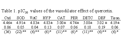
Means ± s.e. means
(n). * P < 0.05; ,** P< 0.01, Students' t test.
In conclusion, quercetin-induced vasodilation is modulated by conditions
of oxidative stress. Drugs which limit quercetin auto-oxidation or drugs
reducing tissue H2O2
formation potentiate quercetin-induced vasodilation. The latter potentiation
operated via an iNOS-dependent mechanism.
Duarte, J. et al., (2001). Br. J. Pharmacol. 133,
117-124.
Hertog, M.G. et al., (1993). Lancet 342, 1007-1011.
Middleton, E. et al., (2000). Pharmacol. Rev. 52, 673-751.