| pA2
online © Copyright 2003 The British Pharmacological Society |
055P
University of Surrey Summer Meeting June 2003 |
|
Evidence that MDMA ('ecstasy') increases interleukin-1ß levels in rat brain
|
Print abstract Search PubMed
for: |
Interleukin-1ß
(IL-1ß) is a pro-inflammatory cytokine that, in the brain, is mainly
produced by activated microglia (Chauvet et al., 2001). IL-1ßis
a key factor in the febrile response to pyrogens (Luheshi et al.,
1999) and a mediator of several forms of brain damage in rodents (Pearson
et al., 1999; Touzani et al., 1999). Administration of 3,4-methylenedioxymethamphetamine
(MDMA or 'ecstasy') to rats produces a long-term degeneration of 5-HT
nerve endings in the brain and an acute hyperthermic response which is
involved in the neuronal damage (Colado et al., 1998).
We have now examined the time-course of changes induced by MDMA on the
concentration of IL-1ß in the cortex and hypothalamus of the rat
and investigated whether these changes are related to the hyperthermic
and neurotoxic responses induced by the drug.
Male Dark Agouti rats (175-200g) received MDMA (12.5 mg kg-1,
i.p.) or saline when housed at a room temperature of 22ºC. Animals
were sacrificed 1 h, 3 h, 6 h, 12 h, 24 h and 7 days later and the levels
of IL-1ß measured by ELISA (Simi et al., 2002). In another
study, rats were maintained at 4ºC, for 4 h before and up to 6 h
after MDMA administration and sacrificed either 3 h or 7 days after drug
injection. Rectal temperature was measured throughout. The 5-HT content
of the cortex was measured by h.p.l.c. 7 days after MDMA injection.
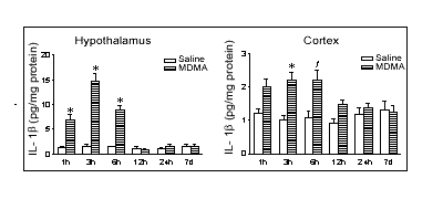
A pronounced increase in IL-1ß levels in the hypothalamus and cortex
was observed 1 h, 3 h and 6 h after MDMA administration with the highest
levels occurring at 3 h in the hypothalamus and at 3 and 6 h in the cortex
(Figure 1). No change was observed 12 h, 24 h and 7 days after MDMA injection
in either brain area. The increase in IL-1ß induced by MDMA at 22ºC
(814% in hypothalamus, 219% in cortex) was significantly reduced, though
not abolished, when rats were kept at 4ºC (454% in hypothalamus,
142% in cortex). In these rats the hyperthermic response was totally abolished
and the long-term loss in cortical 5-HT concentration significantly attenuated.
These findings indicate that immediately after MDMA there is an acute
increase in the brain IL-1ß concentration which is, to some extent,
a consequence of the hyperthermia and suggest that IL-1ß may play
an important role in the neurotoxicity of the drug.

Figure 1. Time-course
of changes in IL-1ß levels in the hypothalamus and cortex induced
by MDMA (12.5 mg kg-1,
i.p.). Results shown as mean ± s.e.mean, n=6-8. Different from
the corresponding saline-treated group: fP<0.01,
*P<0.001.
Chauvet, N. et al., (2001) Eur. J. Neurosci. 14,
609-617.
Colado, M.I. et al., (1998) Br. J. Pharmacol. 124,
479-484.
Luheshi, G.N. et al., (1997) Am. J. Physiol. 272,
R862-868.
Pearson, V.L. et al., (1999) Glia 25, 311-323.
Simi, A. et al., (2002) J. Neurochem. 83, 727-737.
Touzani, O. et al., (1999) J. Neuroimmunol. 100,
203-215.
M.I.C. thanks MCYT (SAF2001-1437) and FIS (02/1885) for financial support.