| pA2
online © Copyright 2003 The British Pharmacological Society |
016P
University
of Manchester Autumn Meeting September 2003 |
| Localisation
of neuropeptide-W 23 (NPW23) and the novel [125I]-NPW-23
ligand to specific nuclei of rat brain
|
Print Abstract Search PubMed
for: |
Neuropeptide-W (NPW) is a recently discovered endogenous ligand proposed for the orphan receptor GPR7 (Shimomura et al., 2002). The mRNA encoding the peptide and receptor protein is expressed in rat brain (Lee et al., 1999; Brezillon et al., 2003). Our aims are to characterise the novel [125I]-NPW-23 ligand to determine if receptors are expressed in native tissues and to localise NPW using immunocytochemistry.
Saturation
binding: For all experiments male Sprague-Dawley rats (350-450g) were
killed using CO2. Binding conditions
were optimised and representative sections throughout the brain were examined
to identify discrete areas of binding for subsequent saturation analysis.
Rat brain sections (10µm) were pre-incubated with 50mM Tris-HCl
buffer containing 5mM EDTA, 10mM MgCl2,
0.3% bovine serum albumin (pH 7.2) for 15 min at room temperature. The
sections were incubated with 7.8pM - 2nM [125I]-NPW-23,
synthesised by Amersham Biosciences (Bucks, UK), in Tris-HCl buffer (as
above) for 1 hr at room temperature. NPW-23 (1µM) determined non-specific
binding. Following a 15 min wash (50mM Tris-HCl, pH 7.4) sections were
apposed to radiation sensitive film for 7 days with [125I]
standards. Specific binding was visualised and determined by means of
computer assisted image analysis (Quantimet 970, Cambridge Instruments).
Data were analysed using the iterative non-linear curve fitting programs
in the KELL package (Biosoft, Cambridge, UK). Pooled KD,
Bmax and Hill slope were expressed as mean ± s.e.mean.
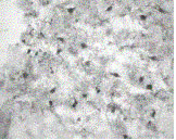
| Figure 1. Presence of NPW-23 (mouse / rat ) immunoreactivity in dorsal raphe nucleus. |
| Immunohistochemistry: Acetone fixed sections were blocked for 1 hr with 5% swine serum. Sections were then incubated with 1:200 rabbit anti NPW-23 primary antibody (Phoenix Pharmaceuticals Inc., CA, U.S.A) for 12 hrs at 4oC. Binding was visualised using the peroxidase anti-peroxidase complex. |
[125I]-NPW-23 bound specifically to the medial amygdala (n = 4) with KD of 0.44 ± 0.13 nM. Receptor density (Bmax) was 149.86 ± 13.83 fmol / mg protein and Hill slopes were close to unity (1.07 ± 0.044). [125I]-NPW-23 binding sites were also localised to the endopiriform nucleus and hypothalamic nucleus. NPW-23 immunoreactivity was observed in the cell bodies of the dorsal raphe nucleus (Figure 1) and ventral tegmental area.
To our knowledge, this is the first evidence for characterisation of [125I]-NPW-23 binding and identification of receptors. The presence of immunoreactive neurones in the dorsal raphe nucleus and ventral tegmental area is consistent with the known projections from these regions to areas of the brain expressing the receptors.
Brezillon
et al., (2003). J Biol Chem; 278, 776-783.
Lee et al., (1999). Mol Brain Res; 71: 96-103.
Shimomura et al., (2002). J Biol Chem; 277, 35826-35832.