| pA2 online © Copyright 2004 The British Pharmacological Society |
068P
GKT, University of London Winter Meeting December 2003 |
|
Premature
endothelial cell replicative senescence in internal mammary artery
endothelial cells cultured from atherosclerotic patients |
|
Cellular senescence is characterised by the limited ability of primary cultured cells to divide. It normally occurs in vivo during the ageing process, but cardiovascular risk factors may lead to premature senescence. To test this hypothesis, we induced replicative senescence in cultured internal mammary artery (hIMA) endothelial cells (ECs) isolated from atherosclerotic patients of various ages.
Segments of hIMA were obtained during coronary artery bypass (n=10, 6 males and 4 females, from 49 to 78 y.o). All patients were dyslipidemic and suffered from angina. 7/10 patients were hypertensive, 5/10 patients were obese and 2/10 were diabetic. ECs were isolated from explants: segments of arteries were placed on Matrigel and incubated in DMEM supplemented with 10% foetal bovine serum, 10% calf serum, 1% penicillin-streptomycin, 90 µg/ml heparin, 100 µg/ml EC growth supplement and 100 U/ml fungizon. Cells were plated onto gelatin-coated culture dishes, characterised by specific immunostaining for ECs and maintained in culture until replicative senescence was reached (from 51 to 103 days of culture). Cytochemical detection of a senescence-associated ß-Galactosidase activity (X-Gal) at pH 6 was used as a marker of senescence. X-Gal positive cells (% of positive cells) were counted at each passage (Dimri et al., 1995).
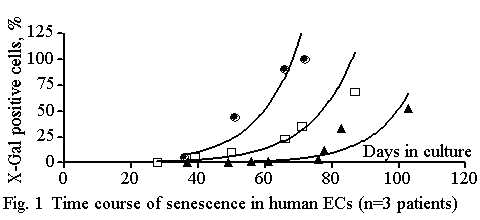
The time course of senescence was exponential (r=0.930) and specific to each patient tested (figure 1). For clarity, data from 3 patients only are shown; From 59 to 100 days in culture (closed circles versus closed triangles) were needed to reach 50% of X-Gal positive cells, suggesting strong heterogeneity between the time courses of senescence in ECs. The slope of the tangents to the curves obtained from each of 10 patients was calculated at the inflexion point by determining the first derivative of the exponential equation. The higher the slope of the tangent, the faster the senescence. Furthermore, as an index of growth, the slope of the linear curves the days in culture and the passage number, was calculated; the higher the slope, the slower the cell growth. This index (x) correlated positively (P=0.0113) with the slope of the tangent (y) (y = 0.406x - 3.027), suggesting that high senescence was associated with a low cell growth rate. In contrast neither senescence nor growth were dependent on the age of the donor (P>0.05).

In conclusion, the dissociation between the age of the donor and the time course of senescence suggests that cardiovascular risks factors lead to premature senescence of the endothelium.
Dimri et al. (1995). Proc Natl Acad Sci USA, 92, 96363-67.