| pA2 online © Copyright 2004 The British Pharmacological Society |
122P
GKT, University of London Winter Meeting December 2003 |
|
Neuroprotective properties of metabotropic glutamate receptor ligands in the 6-hydroxydopamine model of Parkinson's disease
|
|
The cause of dopaminergic cell death in the substantia nigra pars compacta (SNpc) in Parkinson's disease (PD) remains unknown. However, an increasing body of evidence implicates glutamate excitotoxicity in this process (Doble, 1999). Previous studies have suggested that agonists of Group II metabotropic glutamate receptors (mGlu) are neuroprotective in animal models of PD (Murray et al., 2002). We now report the powerful neuroprotective effects of the Group I antagonists, MPEP and LY367385, the Group II agonist 2R,4R-APDC and the Group III agonist, L-AP4 in the rat 6-hydroxydopamine (6-OHDA) model of PD.
Guide cannulae were implanted into the substantia nigra (SN) of male Sprague-Dawley rats (250±20g) under isoflurane anaesthesia, as previously described (Datla et al., 1997). Drug treatment groups (n=3-6) received focal injections of 1) 10nmol MPEP, 2) 200nmol LY367385, 3) 10nmol 2R,4R-APDC or 4) 10nmol L-AP4, or sterile saline alone (4µl). One hour after injection of mGlu receptor ligands the animals were lesioned with 12µg 6-OHDA into the SN. Animals then received daily injections of drug for 7 days; one hour after the final injection the rats were killed by cervical dislocation and the brains rapidly removed. The effects of drug treatment on the integrity of dopaminergic neurons [tyrosine hydroxylase immuno-positive cells (TH+ cells) in the SNpc (Datla et al., 2001)] and the levels of striatal dopamine (DA) (Datla et al., 2001) were then assessed. Data was analysed by ANOVA followed by Student's t-test. 6-OHDA produced a significant loss (61.2±8.9%; P<0.01) of TH+ cells in the control group. The loss of TH+ cells due to 6-OHDA in the animals treated with mGluR ligands was markedly smaller (Figure 1). Similarly, 6-OHDA produced a significant loss (60.33±3.8%; P<0.05) of striatal dopamine in the control group, which was significantly reduced in all drug treated groups (8-20%; P<0.05) comparing the lesioned and unlesioned sides of the brain.
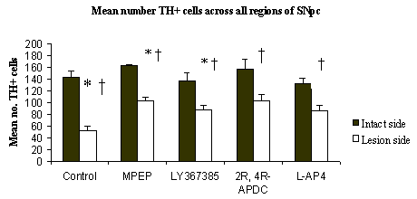
Figure1: Comparison of cell numbers in lesioned vs. intact side (*P<0.01) and cell numbers in lesioned side of control and drug treatment groups (†P<0.01). Data shown are mean cell number ± s.e.m (n=3-6)This study provides compelling evidence for a neuroprotective action of mGlu receptor ligands in the 6-OHDA rodent model of PD.
Datla K.P. et al.
(1997) Neurochemistry. 1157-6.
Datla K.P. et al. (2001) Mov Disord. 16(3), 424-434.
Doble A. (1999) Pharmacol Ther 81(3), 163-221.
Murray T.K. et al. (2002) PharmBiochemBehav.73, 455-466.
A.C.V is the recipient of an MRC PhD studentship.