| pA2 online © Copyright 2004 The British Pharmacological Society |
025P
University of Buckingham 3th Focused Meeting April 2004 |
|
Lateral
hypothalamic neurones which express |
|
The induction of
cGMP by exogenous application of NO-donor compound to brain slices was
investigated in order to identify the targets for NO released in the lateral
hypothalamus.
Wistar rats, either
sex, 15-30 days old were deeply anaesthetised (ketamine HCl 60mg/kg) and
decapitated. The brains were quickly removed and 200µm-thick transverse
slices from the hypothalamic region were cut on a vibrating microtome
in ice-cold, low sodium-artificial cerebral spinal fluid (aCSF), which
was continuously gassed with 95%O2,5%CO2.
The brain slices were then transferred to flasks containing normal aCSF
and slowly warmed up to 37°C in a rocking bath for 15 minutes before
incubation in NO donor, NOC-12, (50 mM) for a further 15 minutes. They
were then fixed and immunostained with anti-cGMP antibody (gift from Dr
J. de Vente). Double immunofluorescent labelling for either orexin-A,
melanin concentrating hormone (MCH), neuronal nitric oxide synthase (nNOS),
glia fibrillary acid protein (GFAP) or the neuronal marker, NeuN, was
carried out.
Brain slices treated with NOC-12 showed cGMP immunoreactivities in GFAP-positive
glia as well as NeuN-positive neurones. The latter were predominant in
the perifonical area, the lateral hypothalamus, and less extensively in
the paraventricular nucleus. Numerous nerve fibres in these areas were
also cGMP-positive. Double immunofluorescent staining for nNOS, orexin
or MCH revealed that none of cGMP-containing neurones expressed these
molecules. In the perifonical area, cGMP-containing neurones appeared
smaller when compared to orexin A-, NOS- and MCH-expressing neurones.
Fig.1 cGMP immunoreactivities in hypothalamus
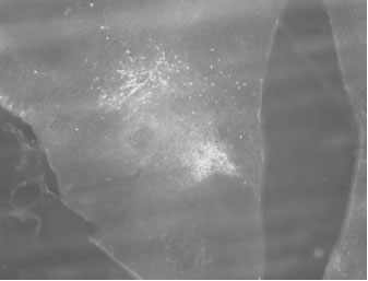
Fig.2. cGMP-expressing neurones in PVN
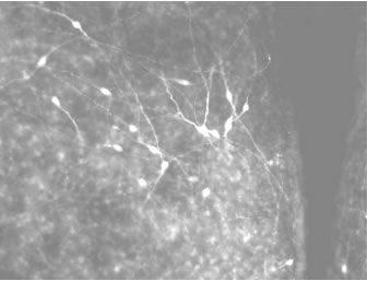
This study has revealed a new population of hyopothalamic neurones, which are neither orexin-, nNOS- nor MCH-containing neurones, and are a potential target for NO released in this region. The cGMP-NO system may play a part in the feeding-sleep regulation (Fetissov et al., 2003: Mariano and Cudeiro, 2003). Further investigation is required for a more comprehensive knowledge of its circuitry and interactions between neurones in this region.
Fetissov et al.(2003).
J. Neuroendoclinol. 15,754-760.
Marino & Cudeiro (2003). J. Neurosci. 23,4299-4307.