| pA2 online © Copyright 2004 The British Pharmacological Society |
032P
University of Bath Summer Meeting July 2004 |
Stimulation of p42,44 mapk by trypsin via PKCε in human cultured prostate stromal cells A. Myatt, DR. Harriss, SJ Hill. Institute of Cell Signalling, Queen’s Medical Centre, Nottingham, UK, NG7 2UH |
|
Trypsin can stimulate p42,44 mitogen-activated protein kinase (MAPK) activity in human cultured prostate stromal cells (Myatt et al., 2003). Here we have investigated the role of protein kinase C (PKC) isoforms in this response to trypsin.
Human cultured prostate stromal cells were grown to confluence in six well plates, serum starved for 2 hours and then stimulated with agonists. Cell lysates were resolved by SDS-PAGE, transferred to nitrocellulose membranes by western blotting and membranes probed with specific antibodies as previously described for measurement of phosphorylated p42,44 MAPK (P-MAPK; Myatt et al., 2003). In some experiments, cells were princubated with phorbol 12,13 dibutyrate (PdBu) for 18 hr prior to agonist stimulation. Where appropriate, PKC inhibitors were added 15 min prior to agonist stimulation. For studies of PKC isoform expression, cells were grown in T75cm2 culture flasks and whole cell lysates of the confluent monolayer resolved by SDS-PAGE, transferred to nitrocellulose membranes and membranes probed with primary rabbit polyclonal antibodies to each isoform (Sigma) and secondary horseradish peroxidase conjugated donkey, anti-rabbit antibody (Amersham). For studies of PKC translocation to the membrane, whole cell lysates were separated into membrane and cytosolic fractions by sonication for 30 seconds and 25000g centrifugation for 20 mins at 4°C and western blotting performed as above. Immunoreactivity was detected with enhanced chemiluminescence. Data were analysed with densitometry (Molecular Analyst software) and statistical significance determined by one-way Anova.
Trypsin stimulation for 15 min caused a concentration- dependent increase in P-MAPK over basal levels (p<0.001; pEC50 7.1 ± 0.3, n=11). Pretreatment of cells with PdBu (18 hr; 1 μM ) completely abrogated the response to trypsin (30nM) compared to vehicle control (p<0.001, n=3). Trypsin (30nM; 15 min) caused significant increases in P-MAPK in the presence or absence of the PKC inhibitor Gö6976 (1μM; p<0.05; n=3). However, a consistent and significant response to trypsin was not obtained in the presence of the PKC inibitors Gö6983 or Ro 318220 (1μM, 10μM respectively). PKC isoforms α, δ, ε were detected in human prostrate stromal cells and could be down-regulated by pre-treatment with PdBu (1 μM ; 18 hr; n=3). PKCζ was detected in these cells but was not down-regulated by PdBu. Trypsin (30nM; 15 min) caused the translocation of PKCε (but not the other isoforms) to the membrane (Fig 1, n=3).
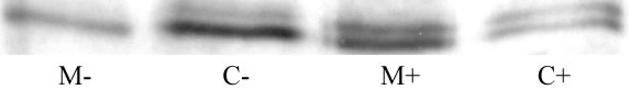
Fig 1. Translocation of PKCε to the membrane by trypsin 30nM. M is membrane fraction, C is cytosolic fraction, +/- is with/without trypsin stimulation respectively. The blot shown is representative of three separate experiments.
These data suggest that trypsin stimulates the phosphorylation of p42,44 MAPK via PKCε in human cultured prostate stromal cells.
Myatt A. et al. (2003). Proceedings of the British Pharmacological Society at http://www.pa2online.org/Vol1Issue2abst019P.html