Print version
Search Pub Med
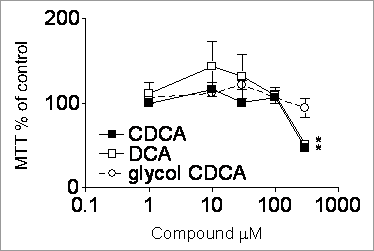
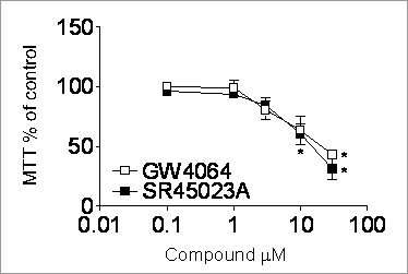
Expression and activation of the farnesoid X receptor in human breast The farnesoid X receptor (FXR; NR1H4) is a member of the steroid super-family of nuclear receptors that acts as a regulator of bile acid synthesis, transport and excretion (Niesor et al., 2001). The plasma bile acid, DCA, is present in high concentrations in breast cyst fluid and in post-menopausal women newly diagnosed with breast cancer (Costarelli et al., 1992). FXR ligands, e.g. CDCA and SR45023A, induce apoptosis in cancerous cell lines including breast carcinoma MCF-7 cells (Niesor et al., 2001; Baker et al., 1992). Thus FXR may represent a novel anti-neoplastic target in breast cancer. Protein and RNA were extracted from cultured MCF-7 and HepG2 cells and 10 paired tumour and next to tumour normal breast tissue samples. The expression of FXR and β actin were assessed by RT-PCR and Western blotting. MCF-7 cells at 60% confluence were growth arrested (no FCS) for 24 hours then incubated for 24 or 48 hours with varying concentrations of bile acids and synthetic FXR ligands (CDCA, DCA, glycol-CDCA, GW4064 or SR45023A) with 0% or 10% FCS. Proliferation was measured by MTT assay and apoptosis by cell and nuclear morphology and ELISA for cytoplasmic histone associated DNA (Bishop-Bailey et al., 2002). MCF-7 cells express FXR mRNA and protein as shown by RT-PCR (Figure 1A) and western blot analysis (Figure 1B), with HepG2 cells being used as a positive control. The bile acids CDCA and DCA (Figure 1C; serum 10%) and synthetic FXR ligands (Figure 1D; serum 0%) caused significant cell death in MCF7 cells. However glycol-CDCA, previously reported to enhance proliferation of MCF-7 cells (Baker et al., 1992), did not cause significant changes in proliferation in this system (Figure 1C). Nuclear condensation and enrichment of cytoplasmic histone associated chromatin fragments indicated the cell death was apoptotic. Both breast tumour and near tumour normal tissue were found to express FXR protein (Figure 1E).
Figure 1 C. Figure 1. Expression of FXR A) RNA and B) protein in MCF7 and HepG2 cells. Effect of C) bile acids CDCA, DCA or glycol CDCA or B) synthetic FXR ligands GW4064 or SR45023A on cell viability. Data represents mean ± SEM of n=3-12; *p<0.05 students t-test, drug compared to control. E) Expression of FXR and β actin proteins in near tumour normal (norm) and tumour human breast samples. In conclusion, our discovery of FXR expression in breast tissue and the fact that FXR ligands induce apoptosis in the breast carcinoma cell line MCF-7 indicates FXR may be a novel therapeutic target in the treatment of human breast cancer. Baker, P. R. et al. (1992) Br. J. Cancer 65, 566-572. This work was funded by the Wellcome Trust (074361/Z/04/Z) and the British Heart Foundation (BS/02/002). |


