5-HT4 receptor distribution in the human adrenal revealed by 3H-GR113808 A 5-HT4 receptor antagonist
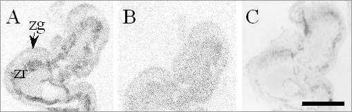
5-HT4 selective agonists stimulate the release of aldosterone but not cortisol in humans (Lefebvre et al. 2002), implying this sub-type is present in adrenal cortex. The aims of this study were to investigate the expression and distribution of 5-HT4 receptors within normal human adrenal and aldosterone-secreting tumours using the 5-HT4 receptor radioligand, 3H-GR113808, previously used to characterise 5-HT4 receptors in the brain (Parker et al, 2003). Human adrenal tissues were obtained, with informed consent, from patients (n=3, 2 female, 1 male, ages 20-42) undergoing unilateral adrenalectomy. Tissues were collected into liquid nitrogen at the time of operation. Cryostat sections of histologically normal regions of adrenal (10 µm) together with the adjacent tumour were cut and thaw-mounted onto gelatin-coated microscope slides and stored at -70 °C until required. Following pre-incubation in Tris-HCl buffer (50mM, pH 7.4) for 15 minutes at 23 °C, sections were incubated with 1nM 3H-GR113808 for 1h, in the absence (total binding) or presence of 10 µM RS-39604 (Parker et al, 2003) to determine non-specific binding. The experiment was terminated by 2x20 sec washes in ice-cold Tris-HCl (50mM, pH 7.4), followed by a final wash (20 sec) in ice-cold de-ionised water. Sections were air-dried and apposed to radiation-sensitive Hyperfilm-3H, with 3H-microscale standards (Amersham Biosciences, GE Healthcare), for 8 weeks; adjacent sections were imaged for 24h using the MicroImager system (Biospace Imaging Systems, France). RT-PCR was performed on RNA from four adrenals. Figure 1. Representative image (MicroImager) showing (A) total 3H-GR113808 binding to zona glomerulosa (zg) and zona reticularis (zr) of normal human adrenal, (B) Non-specific binding. (C) Optical image. Scale bar =5mm.
High levels of specific 3H-GR113808 binding were detected to zona reticularis (Figure 1), of the cortex of normal human adrenal, with lower levels detected in Conn’s tumour, zona glomerulosa and fasciculata. Binding was below the level for detection in the medulla. RT-PCR also showed expression of receptor message in each of the normal adrenals and aldosteronomas (Table 1). Table 1. Specific binding of 3H-GR113808 to normal adrenal and tumour. (mean ± s.e.mean, n=3).
The localisation of receptors to the secretory cells of the glomerulosa is consistent with the observed release of aldosterone in man and the detection of binding in the tumours.
Lefebvre, H., et al. (2002) J. Clin. Endocrinol. Metab., 87, 1211-1216. | ||||||||||||||||