Print version
Search Pub Med

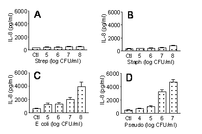
Differential effects of Gram negative versus gram positive bacteria on IL-8 release by human lung epithelial cells: role of toll like receptors 2 and 4 Lung epithelial cells function, not only as a physical barrier, but also as an essential active component of the innate immune system. We now know that innate immunity is regulated by activation of a group of Toll like receptors, 10 of which have been demonstrated in man. TLR2 and TLR4 are the best characterised and are activated by Gram positive and Gram negative bacteria respectively. It is clear that distinct signalling and gene pathways are activated by TLR2 and TLR4, although the extent of the differences are not completely understood. TLR2 forms heterodimers with either TLR1 or TLR6 to mediate functional effects. In the current study we have compared the effects of two types of Gram positive and Gram negative bacteria on IL-8 release by human pulmonary epithelial cells. The human lung epithelial cell line, A549, was cultured to confluence using standard techniques (Mitchell et al., 1997) in 96 well culture plates. Cells were stimulated with increasing concentrations of bacteria (10 4 to 10 8 CFU/ml), LPS (TLR4 ligand; 0.01 to 10µg/ml), Pam 3CSK4 (TLR2/1 ligand; 1 to 1000 ng/ml) or FSL-1 (TLR2/6 ligand; 1 to 1000 ng/ml) for 24 hours. Medium was then removed and IL-8 measured by ELISA.
Figure 1: Effect of increasing densities of bacteria on IL-8 release by A549 cells over 24 hours in culture. A Streptococcus pneumoniae (Strep); B Staphylococcus aureus (Staph); C Escherichia coli (E coli); D Pseudomonas aeruginosa (Pseudo). Data are mean ± S.E. mean for n=8. Gram negative bacteria (Figure 1) or LPS (10 m g/ml) (2574 ± 211pg/ml) but not Gram positive pathogens (Figure 1) increased IL-8 release from A549 cells. Interestingly, by contrast to results with whole bacteria, Pam 3CSK4 (1000ng/ml), which activates TLR2/1 or FSL-1 (1000ng/ml), which activates TLR2/6, increased IL-8 release (3557 ± 276pg/ml and 12067 ± 1048pg/ml respectively). These observations show that TLR4 and TLR2/1 or TLR2/6 activation is positively linked to IL-8 release in human lung epithelial cells. Our findings that whole Gram positive bacteria (which will activate TLR2/1 and TLR2/6 simultaneously) did not induce IL-8 release, suggests that there could be a negative interaction between these pathways, as we have previously demonstrated in vascular cells (Jimenez et al., 2005). Jimenez R, et al. (2005) Proc Natl Acad Sci U S A. 102(12):4637-42. |