150P University of Cambridge
Summer Meeting July 2005 | Cystathionine γ-lyase (CSE) is expressed in human pulmonary cells: relevance to pulmonary hypertension R. Faro, G. Sturton , G. Cirino* and J.A.Mitchell. Cardiothoracic Pharmacology, Unit of Critical Care, National Heart and Lung Institute, Imperial College, London, UK and *Dipartimento di Farmacologia Sperimentale, Universita di Napoli-Federico II, 80131 Naples, Italy. Nitric oxide (NO) and carbon monoxide (CO) are simple gases that easily pass through biological membranes. They are characterised by continuous generation, fast transmission, and extensive action. NO and CO have been implicated in the regulation of pulmonary vascular homeostasis and a reduction in the endogenous production of these dilator gases has been associated with pulmonary hypertension. Hydrogen sulfide (H2S) is now similarly recognised as an endogenously generated vasodilator gas. Endo genous H2S can be formed from cysteine by pyridoxal-5’-phosphate-dependent enzymes, including cystathionine β-synthase (CBS) or cystathionine γ-lyase (CSE). The aim of this study was to characterise the expression of CSE in human pulmonary vascular tissue and to demonstrate the action of H2S on human on vasomotor responses in human lung vessels. Confluent human pulmonary artery smooth muscle (HPASM) and endothelial (HPAEC) cells (passage 2–6), seeded on 6-well plates were treated with inflammatory stimuli; Escherichia coli (E.coli) or Staphylococcus aureus (S.aureus) in presence or absence of human recombinant IL 1β (10 ng/ml) for 24 h, after which, cell supernatant was discarded and cells were scraped into gel loading buffer and extracts boiled for 20 min. Western blot analysis was performed using standard techniques with anti-CSE-antibody (1/1000), kindly provided by Dr. Ishii - Department of Molecular Genetics – Tokyo –Japan. Consistent protein concentrations were estimated byPonceau S staining. Figure – Western Blot for cystathionine γ-lyase (CSE) in H uman pulmonary artery smooth muscle (HPASM) and endothelial (HPAEC) cells. 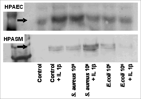
CSE was expressed constitutively in HPAEC. Lower levels were detected in HPASM, however, levels were increased by IL-1, S.aureus or E.coli. H2S (delivered by solutions of sodium hydrogen sulphide (NaHS; 0.1-30µM) induced vasodilation of U-46619 constricted human pulmonary vessels (34.4 + 5.1% of induced tone - for NaHS 10µM), measured visually using precision-cut lung slices ( Parrish et al., 1995 ) . These observations show CSE is expressed in human pulmonary vascular cells and are the first to demonstrate that this enzyme can be regulated by inflammatory insult. Our data illustrate the potential role of the CSE – H2S pathway in pulmonary hypertension. Parrish AR et al. , Life Sciences 57: 1887–1901, 1995. | 
