Effect of prolonged exposure to tumour necrosis factor-alpha and nicotine on rat mesenteric and pulmonary arteries
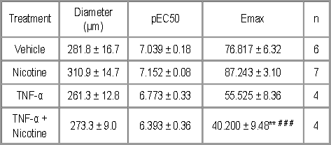
The pro-inflammatory cytokine tumour necrosis factor-α (TNF-α ) is associated with various vascular disorders including atherosclerosis and peripheral arterial disease. It has previously been reported that acute exposure to TNF-α inhibits endothelium-dependent relaxation in rat mesenteric arteries (Wimalasundera et al., 2003). Nicotine, as a constituent of cigarette smoke, has been identified as a major contributor to vascular disease and has been shown to impair endothelial-dependent relaxation in porcine carotid and coronary arteries (Conklin et al., 2001). In the present study, we have examined the effects of nicotine exposure on endothelial-dependent relaxation in pulmonary and mesenteric arteries, in the presence of TNF-α , a mediator produced by a variety of cells, including endothelial cells, under conditions of inflammation. Male Wistar rats (240-260g) were sacrificed by CO2 asphyxiation. Mesenteric and pulmonary arteries were dissected and treated with 6-hydroxydopamine (2 mM) and capsaicin (0.1 mM) for 30 minutes in order to remove neuronal influences. Following this, some vessels were incubated in DMEM containing nicotine (10-7 M) and TNF-α (10 ng ml-1), alone and in combination, or vehicle, for a period of 24 hours in respect of pulmonary arteries and 48 hours for mesenteric arteries. 2 mm segments of artery were mounted on a wire myograph under normalised tension in oxygenated (95% O2/5% CO2) Krebs’ buffer maintained at 37°C. Maximum contraction to KCl (120 mM) was initially determined and sub maximal tone subsequently induced using phenylephrine (0.1-10 µM) or U46619 (0.01-1 µM) in the presence of nifedipine (0.3 µM). Endothelial-dependent responses to acetylcholine (ACh) were used as a measure of endothelial function. Maximal responses to ACh (Emax) are expressed as percent relaxation of active tone (mean ± SEM) and differences in pEC50 and Emax determined by ANOVA followed by Bonferroni’s post test. Table 1 Pulmonary arteries (48 hour incubation)
Table 2 Mesenteric arteries (24 hour incubation) ** P<0.01, *** P<0.001, denotes difference between vehicle control and treated vessels. Pulmonary arteries exposed to TNF-α for a period of 24 hours showed significantly reduced maximal endothelial-dependent relaxation in response to ACh (Table 1). Interestingly, mesenteric arteries treated with TNF-α alone for 48 hours did not differ significantly from vehicle treated vessels, however, TNF-α exposure in the presence of nicotine did significantly reduce subsequent endothelial-dependent relaxation (Table 2).
Conklin et al. (2001). J. Surg. Research. 95 23-31. |


.gif)
