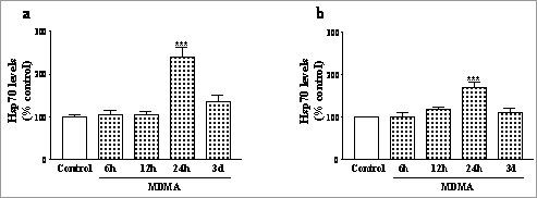
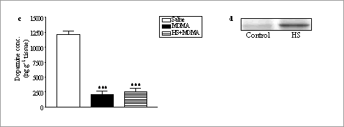
MDMA increases Hsp70 in mouse striatum and hippocampus but heat shock induced Hsp70 does not protect against dopamine depletion Heat shock protein 70 (Hsp70) expression is increased following hyperthermia (heat shock, HS, Leoni et al., 2000) and is thought to be involved in neuroprotection. 3,4-Methylenedioxymethamphetamine (MDMA, ‘ecstasy’) produces a long-term reduction in striatal dopamine in mice which is modulated by the acute hyperthermic response produced by the drug (Green et al., 2003). We have now studied the time-course of MDMA-induced expression of Hsp70 and explored if prior induction of this protein by HS protects against MDMA-induced dopamine (DA) toxicity. Adult male C57BL/6J mice (25-30 g) were given MDMA (30 mg kg-1, i.p. x 3, 3 h intervals) and killed 6, 12, 24 h or 3 d after the last dose. Hsp70 expression in striatum, hippocampus and cerebral cortex was detected by Western blot and levels in striatum and hippocampus quantified by an enzyme-linked immunosorbant assay. A separate group of mice was anaesthetised with pentobarbitone (40 mg/kg., i.p.) and placed in a cage maintained at 38ºC by immersion in a water-bath to raise their rectal temperature to 42ºC for 10 min (HS). Twenty-four h later these mice were either killed for the determination of Hsp70 expression or given MDMA (30 mg kg-1, i.p. x 3, 3 h intervals) at an ambient temperature of 21ºC and rectal temperature monitored for 7 h. Animals were killed 7 d after MDMA and striatal DA determined by h.p.l.c. MDMA produced a significant increase in Hsp70 expression in the mouse striatum and hippocampus 24 h after drug administration (Fig. 1 a, b). No effect was observed in the cerebral cortex at any of the times studied. HS pretreatment significantly increased Hsp70 expression in the striatum 24 h later (Fig. 1d) but did not protect against the MDMA-induced reduction in striatal DA levels 7 d later (Fig. 1c). MDMA produces a region-specific and time-dependent increase in Hsp70 levels but over-expression does not protect against the neurotoxicity induced by the drug.
Figure 1. Time-course of Hsp70 protein levels after MDMA (30 mg kg-1) in (a) striatum and (b) hippocampus. (c) Striatal DA levels 7 d after treatment with MDMA with and without prior HS and (d) representative Western blot of HS-induced Hsp70 expression in striatum 24 h after exposure compared with controls. Results shown as mean ± s.e.mean, n=3-7. Different from controls: ***P<0.001 (Tukey’s test). Green, A.R. et al., (2003) Pharmacol. Rev. 55, 463-508. E.O.S. thanks CAM ( 08.8/0004.1/2003), PNSD (MSC) and MCYT ( SAF2003-05180) for support. |


