Print version
Search Pub Med
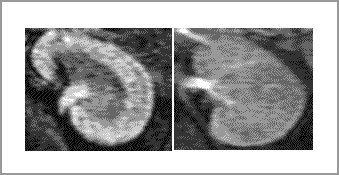
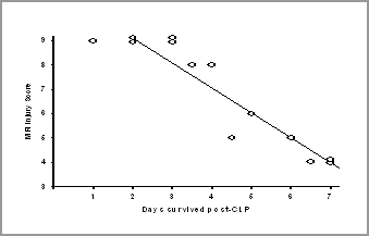
In vivo detection of renal injury and drug action using a novel MRI technique in a mouse model of sepsis The objective of this study was to develop a non-invasive method based on magnetic resonance imaging (‘dendrimer-enhanced MRI’) 1 which could detect a drug effect in vivo in a mouse model of sepsis-induced acute renal failure (ARF). We also investigated whether dendrimer-enhanced MRI could distinguish drug-induced ARF from sepsis and whether imaging could provide prognostic information. We performed MRI with gadolinium-based G4 dendrimer intravenous contrast 2 . We used a fluid- and antibiotic- treated cecal ligation and puncture (CLP) sepsis model in aged,male, C57BL mice (approved by the NIH ethics committee). We have previously demonstrated that the anti-inflammatory agent ethyl pyruvate (EP) can prevent organ injury in this model 3 . Therefore, we monitored the effect of EP on renal function with MRI. Twenty hours post-CLP, aged mice had a distinct pattern of renal injury using dendrimer-enhanced MRI (figure 1). This pattern was different from renal injury induced by cisplatin nephrotoxicity. EP reversed the renal dysfunction even when administered after renal injury was detected by MRI. Dendrimer-enhanced MRI detected renal dysfunction 6 hours post-CLP, a time when serum creatinine was still normal. The appearance of renal dysfunction on dendrimer-enhanced MRI at 6 hours post-CLP predicted the length of survival (fig 1). Dendrimer-enhanced MRI is a novel biomarker that provides information for the early diagnosis, drug responsiveness and prognosis of sepsis-induced ARF.
Normal Sepsis
FIGURE 1 – Sepsis produces a distinct pattern of renal injury compared with normal. The severity of this pattern at 6 hours post-CLP predicts survival 1. Dear JW, Kobayashi H, Jo SK et al. Dendrimer-enhanced MRI as a diagnostic |
|


