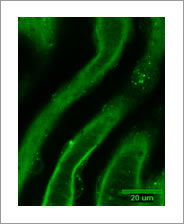
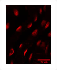
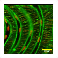
Visualisation of endothelial α1-adrenoceptors in the murine carotid artery The presence of α1 -adrenoceptors (AR) on the vascular endothelium is controversial. However, the first functional evidence of endothelial α1- ARs was recently reported (Fillipi et al., 2001). This confocal imaging study of the murine carotid artery used Quinazoline Piperazine Bodipy (QAPB; Molecular Probes Inc., Eugene), a fluorescent analogue of prazosin, and Syto 61, a live nuclear dye, to visualise endothelial α1-ARs. C57 Black mice (4 months; 29.2g-40.4g) were killed by a lethal overdose of carbon dioxide. Carotid arteries were removed and incubated (21°C, 120 minutes) with either 0.1μM QAPB and 1μM Syto 61, or a solution containing 0.1μM QAPB, 1μM Syto 61 and 0.1μM prazosin (a nonselective a 1-AR antagonist) . All compounds were diluted in PSS and incubation solutions were replaced every 30 minutes to ensure that pH was maintained. Arteries were sliced open with a single-edged razor blade, enabling the vessel to be mounted endothelial side up on a glass microscope slide with a cover slip on top (Miquel et al., 2005). All arteries were visualised using the Bio-Rad Radiance 2100 Confocal Laser Scanning System. The excitation/ emission wavelengths used were 515nm/ 530nm for QAPB and 637nm/ 660nm for Syto 61 (x40 objective). When comparing images the same laser intensity and gain were selected. An image size of 515 x 512 pixels produced a field size of 289μm x 289μm. Five-framed Kalman were used to record images and five random areas of artery were imaged in each mouse (n=6). Endothelial cells (Figures 1 & 2) were clearly identified by their position and nucleus shape using Syto 61. QAPB binding to endothelial cells was diffuse and clustered binding was observed mainly on the surface of the cell membrane. In the presence of prazosin, no QAPB binding to endothelial cells was identified. In conclusion, we have provided further evidence in support of endothelial α1- ARs in the mouse vasculature.
Figure 1. Binding to endothelial cells on the internal elastic lamina. Green panel shows clustered QAPB binding (laser 40, gain 12) and red panel shows Syto 61 nuclear binding (laser 30, gain 30).
Figure 2. Overlay of QAPB (green: laser 40, gain 12) and Syto 61 (red: laser30, gain 30).
Fillipi, S. et al. (2001) JPET, 296: 869-875. |
|