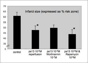
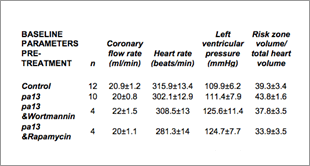
Pyr1-apelin-13 reduces myocardial reperfusion injury independent of PI3K/AKT and P70S6 kinase Apelin peptides are the endogenous ligands of the former orphan G-protein coupled apelin receptor (previously known as APJ). Apelins are predominantly expressed in vascular endothelial cells and adipocytes and regulate vascular tone, cardiac contractility and more recently have been proposed to protect from isoprenaline-induced myocardial injury (Kleinz et al., 2005). Furthermore, transgenic cell-based assays suggest that apelin peptides activate two major components of the reperfusion injury salvage kinase (RISK) pathway, PI3K/Akt and P70S6 kinase (P70S6K), a pathway which mediates the cardioprotective effects of many agents that limit myocardial ischaemia/reperfusion injury (Hausenloy et al., 2006). Based on these findings we hypothesised that apelin peptides may protect the post-ischaemic myocardium from reperfusion injury via the RISK pathway and tested this hypothesis in a rat isolated heart model of ischaemia and reperfusion. Male Sprague-Dawley rats (290-350g) were anaesthetised with pentobarbital sodium (60mg/kg i.p.) and anticoagulation was achieved by coadministration of heparin (1000IU/kg). Hearts were excised and retrogradely perfused via the ascending aorta at 11.3 kPa with oxygenated (95%O2/5%CO2) Krebs’ buffer at 37°C. A 3-0 suture held by a snare was placed around the left main coronary artery (CA) to allow reversible regional ischaemia. After 20 minutes of equilibration the coronary artery was occluded for 35 min, followed by 120 min of reperfusion. Ischaemic risk zone was measured by Evans’ blue infusion after reocclusion of the CA. Infarct size was determined by triphenyltetrazolium staining and expressed as % risk zone. Pyr1-apelin-13 (10-8M) was administered for 20 min starting 5 min before onset of reperfusion. To investigate a possible involvement of PI3K/Akt or P70S6K in mediating Pyr1-apelin-13 actions, the same protocols were carried out in hearts treated with wortmannin (10-7M) and rapamycin (10-9M) starting 10 minutes before Pyr1-apelin-13 treatment for 30 minutes. Pyr1-apelin-13 (pa13) reduced infarct size by 22% compared to controls (Figure 1). This was not abolished by pretreatment of hearts with wortmannin or rapamycin.
Figure 1: Data are mean ±SEM. *indicates statistically significant difference to control (<P0.05) determined by one-way ANOVA in combination with Dunnet’s post-hoc test.
Our results indicate that in line with the expression of apelin receptors on cardiac myocytes, Pyr1-apelin-13 reduces reperfusion injury in the post-ischaemic heart. The PI3K/Akt and P70S6K inhibitors wortmannin and rapamycin did not abolish apelin cardioprotection, suggesting involvement of alternative pathways in native tissues. Kleinz et al. (2005) Pharmacol Ther. 107(2):198-211. |
|


