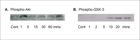
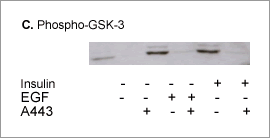
Akt inhibitor A-443654 shows Akt is necessary for epidermal growth factor stimulation of hepatocyte proliferation Epidermal growth factor (EGF) enhances survival and cell cycle progression of cultured primary hepatocytes in a manner dependent on the phosphoinositide 3-kinase / Akt / mammalian target of rapamycin (PI3K/Akt/mTOR) pathway (Coutant et al., 2002; Schulze-Bergkamen et al., 2004), and yet it has been reported that dominant-negative Akt does not inhibit EGF-stimulated DNA synthesis (Coutant et al., 2002) . Here have used for the first time a chemical inhibitor of Akt, A-443654 (see Luo et al, 2005 for structure) to more directly investigate the role of Akt in proliferative responses to EGF in primary hepatocytes. Rat hepatocytes were cultured for 4h in the presence of insulin (10 μg ml -1) and foetal calf serum. After a further 20h culture in the absence of both serum and insulin EGF (10 μg ml -1) was added as indicated. [3H]thymidine incorporation into DNA and MTT ( (3-(4,5-Dimethylthiazol-2-yl)-2,5-diphenyltetrazolium bromide)) assays were undertaken 24h later. A-443654 (3 μM) was added 30 mins prior to EGF, as appropriate, and at times indicated the medium was replaced with inhibitor free medium with EGF, so when present EGF was always for 24h. Western blot for phospho-Akt and phospho-glucose synthase kinase 3 (GSK-3) (a substrate of Akt) were used as an index of Akt phosphorylation and activity respectively. EGF stimulated [3H]thymidine incorporation into DNA (control 78.9 ± 3.5, EGF 158.3 ± 9.0, d.p.m. x 10-3, mean ± s.e.mean, n = 4, P<0.001 Students t-test). When A-443654 was present for 1h or more of the 24h EGF stimulation, the response was lost. (1h inhibitor: control 35.6 ± 3.1, EGF 35.7 ± 7.8. 2h inhibitor: control 33.2 ± 2.2, EGF, 37.3 ± 0.7. 4h inhibitor: control 28.1 ± 1.5, EGF, 27.6 ± 6.8; d.p.m. x 10-3, mean ± s.e. mean, n = 4). When an MTT assay was undertaken on parallel cultures to indicate the number of viable cells there was no substantial loss in response to the presence of A-443654 at any of these times (not shown). Data are not representative of 3 separate experiments.
Figure 1. Western blots for phospho-Akt and phospho-GSK as indicated. In A and B EGF was present for the times indicated; C, insulin (10 ug ml-1) and EGF were present for 5 mins, with A-443654 (A443) added 30 mins prior to stimulations. These results show: 1. EGF stimulates cell cycle progression, Akt phosphorylation and Akt activity in primary rat hepatocytes; 2. the Akt inhibitor A-443654 is able to block both EGF-stimulated Akt activity and cell cycle progression. The results support an essential role for Akt in the proliferative response to EGF in hepatocytes.
Coutant, A et al (2002) Hepatology 36, 1079-1088. |
|