091P Brighton
Winter Meeting December 2008 |
Human IL-15/IL-15Rα fusion protein mediates an increase in NK1.1 spleen cells in the mouse
Anita Midha1, Jaimini Reens1, Anne Mathers1, Robert Craggs1, Alison Ward1, Philip Mallinder1, Donna Finch2, Matthew Sleeman2, Simon Cruwys1, Philip Kerry1
1AstraZeneca R&D Charnwood, Loughborough, East Midlands, UK, 2MedImmune Ltd, Cambridge, UK
IL-15 is a cytokine involved in the development of NK cells. In vivo IL-15 is normally presented in a cell-associated form bound to its high affinity receptor IL-15Rα. The biological activity of soluble IL-15 is enhanced by soluble IL-15Rα. When administered to mice, recombinant IL-15 induces splenomegaly with an associated increase in splenic NK1.1 cells. Administration of IL-15/ IL-15Rα complex gives an enhanced response suggestive of a superagonist effect (Rubinstein, et al, 2006). We have further characterised the biological effects of soluble IL-15/IL-15Rα complex in mice using a surrogate fusion protein of human IL-15Rα sushi domain fused to IL-15 (IL-15/IL-15Rα fusion protein).
C57BL/6/J male mice (5/group) were dosed i.p. with PBS, human IL-15 (hIL-15) or IL-15/IL-15Rα fusion protein once daily for 3 days. Single cell suspensions were prepared from half the spleen and percentage NK1.1 positive cells were determined by flow cytometry. Histological sections of remaining spleen tissue and the sternums, were stained with haematoxylin & eosin (H & E)
IL-15/IL-15Rα fusion protein induced an increased spleen weight. This splenomegaly was associated with expansion of the red pulp, with increased numbers of progenitor cells, suggestive of haematopoiesis. IL-15/IL-15Rα fusion protein induced leucocytosis in peripheral blood, with increased numbers of large atypical lymphocyte-like cells, a subpopulation of which was shown, by immunohistochemistry and flow cytometry, to be NK1.1 positive cells. Flow cytometry analysis of spleen cell suspensions from these treated mice also had increased numbers of NK1.1 cells (Figure 1). IL-15/IL-15Rα fusion protein -treated mice also exhibited an increase in the M:E ratio (myeloid hyperplasia) in sternal bone marrow.
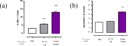
Figure 1. (a) NK 1.1 Expression on C57BL/6/J Mouse Spleen Cells, (b) Spleen weights relative to body weights
These data demonstrate that IL-15/IL-15Rα fusion protein not only mediates an increase in NK1.1 cells but also causes an increase in haematopoietic activity. This model may be of use to further investigate IL-15 as a potential disease target.
Rubinstein, MP, et al. Proc Natl Acad Sci . 2006, 103:9166-71.
|

