
Up-regulation of interleukin-10 by adrenomedullin in rat alveolar macrophage and human THP-1 monocytic cell lines: role of the p38-mitogen-activated protein kinase and protein tyrosine kinases Adrenomedullin (AM) is a potent vasorelaxant peptide that plays a role in inflammation. Transgenic mice over-expressing AM are resistant to septicaemic shock. Lipopolysaccharide (LPS) induces proteins involved in the inflammatory response, including mitogen-activated protein kinases (MAPKs) and protein tyrosine kinases (PTKs). We investigated the effect of AM on interlukin-10 (IL-10) production in NR8383 rat alveolar macrophage and THP-1 human monocytic cell lines (American Type Culture Collection, Rockville, MD) and the regulatory pathways involved. AM augmented significantly LPS-induced IL-10 production in both NR8383 cells and phorbol 12-myristate 13-acetate (PMA)-activated THP-1 cells by 66.8% (p<0.01) and 66.4% (P<0.05) respectively (Fig.). In contrast, interferon-γ (IFN-γ) markedly inhibited LPS-induced IL-10 production in both cell types by 81.5% (p<0.05) and 77.4% (p<0.01) respectively. In NR8383 cells, LPS-induced IL-10 production was reduced from 41.4±5.9 (mean±SEM) to 18.3±2.2 pg/ml (p<0.01) by SB203580, an inhibitor of p38-MAPK, whereas inhibition of p42/44-MAPK had no effect. Inhibiting IL-10 production in NR8383 cells through blocking p38-MAPK was partially rescued by adding exogenous AM and interleukin-6 (IL-6) but not tumour necrosis-α (TNF-α). We also found that the IL-10 production in NR8383 cells was reduced from 259.1±22.7 to 35.0±6.7 pg/ml by 0.1 mM genistein (p<0.001), a protein tyrosine kinase inhibitor, and the inhibition could not be rescued by AM, IL-6 or TNF-α. Our results suggest that LPS-stimulated IL-10 production in macrophages may be regulated by AM via pathways which depend on p38-MAPK and PTKs. These findings provide further evidence that AM plays a role in the regulation of inflammation. 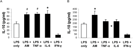
Fig. Effect of AM and inflammatory mediators on LPS-induced IL-10 production in NR8383 and PMA-activated THP-1 cells. A, IL-10 concentration at 24 hr after stimulation with 1 ng/ml LPS in the presence or absence of other mediators in NR8383 cells. B, IL-10 concentration at 24 hr after stimulation with 1 ng/ml LPS in the presence or absence of other mediators in PMA-activated THP-1 cells. Data are mean ± SEM of 4-6 separate sets of experiment. *, P<0.05; #, P<0.01 vs. LPS only. |
|
