138P Brighton
Winter Meeting December 2008 |
Effect of pre-ischaemic conditioning on hypoxic depolarization of dopamine efflux in the rat caudate brain slice measured in real-time with fast cyclic voltammetry
Colin Davidson, Ben Coomber, Claire Gibson, Jonathon Stamford, Andrew Young
Leicester University, Leicester, UK
Pre-ischaemic conditioning (PIC) with brief periods of ischaemia has been shown to be neuroprotective against a subsequent ischaemic event. Here we examined whether short periods of PIC can affect hypoxia induced dopamine (DA) release. We have found that by switching the perfusion medium from oxygenated to deoxygenated solutions we can evoke high levels of DA release from caudate brain slices (Toner & Stamford, 1996). The extent to which released DA is involved in neurotoxicity is debated.
Male Sprague-Dawley rats (∼250 g) were killed by cervical dislocation and their brains rapidly removed. Brain slices (400 μm) were kept in oxygenated artificial cerebrospinal fluid (aCSF) at room temperature (22°C). After at least 45 min equilibration, slices were transferred to a brain slice chamber and perfused with oxygenated aCSF at 35°C. After a further 45 min equilibration in the slice chamber, they were subject to PIC (0, 2 or 10 min) comprising hypoxia (N2/CO2) and reduced glucose (10 to 2 mM) aCSF, then, 60 min later, a 15 min hypoxic episode. DA was measured using fast cyclic voltammetry (FCV) at carbon fibre electrodes as previously described (Toner & Stamford, 1996). Electrodes were calibrated in DA. We recorded a) time from start of hypoxia to start of DA release (t-on); b) time from start of DA release to peak DA release (t-peak); c) peak DA release and d) initial rate of DA release (dDA/dt). Data were analyzed with 1-way ANOVA.
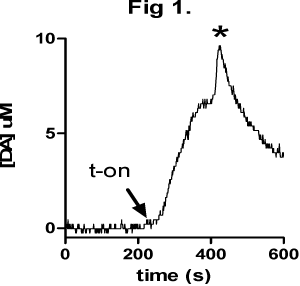
There was no hypoxia-induced DA release after 0 or 2 min PIC, however 10 min PIC evoked DA release after ∼300 s (Table 1 “none”). Occasionally we observed a secondary fast DA peak (Figure 1*). 10 min PIC decreased the amount of DA released (p<0.05) and also the rate of DA release versus 0 min PIC (p<0.05). There was a tendency for 10 min PIC to increase t-on, the time to DA release after hypoxia.
These data suggest that PIC may have a neuroprotective effect in caudate slices; the time of onset of hypoxia-induced DA release tended to be increased after 10 min PIC, while the same length of PIC decreased the amount of DA released and the rate of DA released on a subsequent hypoxic event. It is debatable whether the reduced rate or reduced amount of DA release seen after 10 min PIC are due to a neuroprotective effect of PIC, or indeed whether these are a sign of ill-health. Correlation with other measures of tissue health are needed (Mathews et al., 2000).
Mathews KS et al., (2000) J Neurosci Meth 102, 43-51.
Toner CC and Stamford JA (1996) J Neurosci Meth 67, 133-140

Table 1. means ± SEM
| PIC (min) | n | t-on (s) | t-peak (s) | [DA] (μM) | dDA/dt (nM/s) |
| 0 |
4 |
271±20 |
115±7 |
8.4±1.0 |
75±10 |
| 2 |
4 |
252±40 |
153±27 |
7.0±1.0 |
51±13 |
| 10 |
4 |
372±69 |
156±21 |
3.5±0.5* |
26±5* |
| none |
4 |
297±56 |
94±13 |
10.3±1.7 |
113±22 |
|


