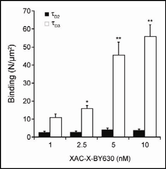
Using the fluorescent antagonist, XAC-X-BY630 to quantify antagonist-adenosine A3 receptor complexes in membrane microdomains of single living cells. The adenosine A3 (A3-AR) receptor is one of four known G-protein coupled receptors activated by the nucleoside adenosine. We have previously demonstrated the heterogeneity of agonist-A3-AR complexes in the membrane of Chinese Hamster Ovary (CHO-A3) cells using fluorescence correlation spectroscopy (FCS) (Cordeaux et al., 2008). Here, we describe the characterisation of XAC-X-BY630 (Briddon et al., 2004), as a fluorescent antagonist label for the A3-AR and use it to demonstrate a similar heterogeneity in antagonist-A3-AR complexes in CHO-A3 cells. Confocal imaging and FCS were carried out essentially as described (Cordeaux et al., 2004). Confocal imaging showed clear membrane labelling of CHO cells expressing a YFP-tagged version of the A3-AR following incubation with XAC-X-BY630 (25nM, 10min, 22°C), which was substantially reduced by MRS1220 (100nM, 30min, 37°C) (n = 3). Initial FCS experiments using CHO-A3 cells incubated with XAC-X-BY630 (1-10nM, 10min, 22°C) revealed both fast- (τD2) and slow-moving (τD3) complexes at the cell membrane, with average diffusion co-efficients of 1.58±0.16 μm2/s and 0.081±0.007 μm2/s, respectively (mean±s.e.mean, n = 100). At concentrations of XAC-X-BY630 ranging from 1-10nM the amount of τD3, but not τD2, increased in a concentration-dependent manner (Fig.1 P<.0001, one-way ANOVA). However, both species represent antagonist-receptor complexes, since the binding of both τD2 and τD3 following addition of 2.5 and 5nM XAC-X-BY630 was significantly reduced following pre-incubation of cells with the specific A3-AR antagonist MRS1220 (300nM, 30min, 37°C) (P<0.01, t-test, n = 12). Fig.1 Quantification of XAC-X-BY630 binding in CHO-A3 cells using FCS. CHO-A3 cells were incubated with XAC-X-BY630 (10min, 22°C) and levels of τD2 and τD3 determined. mean±s.e.mean, n = 34-48. *P<0.05, **P<0.001 vs 1nM, Newman-Keuls test. 
Cordeaux, Y et al. (2008) FASEB. J., 22, 850. We thank MRC and BPS for financial support. |
|

