Effect of chronic stress and antidepressant treatment on rat brain mitochondrial reactive oxygen species (ROS) production Hypercortisolism is known to underlie depressive illness; in fact depression is thought to be a behavioural representation of activated primary mediators of the stress response (Conti et al., 2004). Therefore over the years a number studies have sought to learn the effect of antidepressant treatment on serum corticosterone profile. This study aimed to take this a step further and explore the relationship between serum corticosterone, antidepressants and mitochondrial function. Treatments lasted 14 days using a 2 × 2 factorial design, where animals received 10mg kg-1 fluoxetine or 10mg kg-1 imipramine, intra-peritoneally, once a day at approximately 10:30am. Control animals were injected with vehicle. Concurrently animals received corticosterone (CORT; 5mg L-1) or its vehicle ethanol (EtOH; 0.5% vv-1) orally through drinking water. Trunk blood was collected at the end of the 14 day period. Forebrain mitochondria were isolated as previously described (Markham et al., 2004). Serum corticosterone concentration was determined using a quantitative enzyme immunoassay. Reactive oxygen species (ROS) production was measured fluorometrically as described previously (Markham et al., 2008).

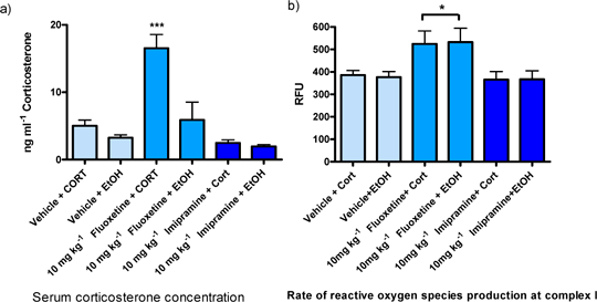
Figure 1 The effect of antidepressants and oral corticosterone on a) circulating serum CORT and b) mitochondrial ROS production, expressed as relative fluorescence units. The data were analysed using one-way ANOVA followed by Tukey’s post-test. Bars represent mean ± s.e.m.; n = 3; *P<0.05 and ***P<0.001, compared to rest.
Animals receiving fluoxetine in combination with oral corticosterone had significantly (P<0.001) higher concentrations of serum corticosterone, compared to all other groups. Additionally, fluoxetine treated animals had a significantly (P<0.05) higher rate of ROS production, regardless of whether they received oral CORT or EtOH, compared to those treated with imipramine and control animals. This effect was specific to complex I supported respiration. The decrease in serum corticosterone after treatment with imipramine is a part of the mechanism of action of this drug. Imipramine corrects the suppression of the HPA feedback loop that is associated with stress (Piwowarska et al., 2009). While the increased levels of circulating serum corticosterone following fluoxetine injections can be explained by the ability of fluoxetine to act as an inducer of serum corticosterone for up to 2 h after administration (Yirimia et al., 2001). Therefore, the timing of sample collection is critical in chronic studies. This study is unique in that no one has previously studied or shown the effect of concurrent in vivo treatment with antidepressants and corticosterone on rat brain mitochondrial ROS production.
Conti et al., (2004) J. Nuerosci. 24(8), 1967-1975 Markham et al., (2004). Eur. J. Neurosci. 20, 1189-119 Markham et al., (2008) Proc. B.P.S. 5(2)-053P at http://www.pA2online.org/abstracts/Vol5Issue2abst053P.pdf Piwowarska et al., (2009) Pharmacol. Reports, 61, 604-611 Yirimia et al., (2001) Neuro. Psych. Pharam. 24 (5), 531-544
|
|

