
Print version
Search Pub Med

Steroids only partially affect polyinosinic:polycytidylic acid (Poly I:C)-induced airway inflammation and hyper-responsiveness in mice Background: Poly I:C is a synthetic dsRNA used to model viral infections and exacerbations of chronic respiratory diseases (e.g. asthma and COPD). There are no thorough in vivo characterizations of the inflammatory and physiological changes induced by a single administration of poly I:C, a TLR3 agonist, in the literature. Methods: Male BALB/C mice (12 weeks) were intranasally administered poly I:C under short-acting anaesthetic. To determine if airway hyper-responsiveness (AHR) was a feature of the model, mice were challenged with increasing doses of nebulized methacholine (MCh) (1-300 mg/ml). Subsequently, bronchoalveolar lavage (BAL) was performed and lung tissue was digested or fixed with 10% NBF to evaluate changes in inflammatory cell infiltrate. For studies evaluating steroid, budesonide (0.3-10 mg/kg) or vehicle (2% Klucel, 0.1% Tween 80 in water) was administered per os 1 hour prior to poly I:C challenge and AHR and inflammation were assessed 24 hours later. Results: Administration of poly I:C (3 – 100 µg) induced dose-dependent increases in neutrophils and lymphocytes in the BAL fluid. A submaximal dose of 30µg was used for subsequent studies. A time course study revealed neutrophils and lymphocytes increased in the BAL fluid between 6 and 72 hours after poly I:C administration before resolving back to baseline at 7 days. Analysis of these BAL fluid cells by flow cytometry revealed the increased numbers of lymphocytes were largely NK and CD8+ T cells, whereas there was no change in the CD4+ population. Poly I:C-treated mice also had enhanced airway responses to cholinergic challenges 24 hours post-administration as indicated by a drop in the PC100 value (concentration of MCh needed to increase pulmonary insufflation pressure (PIP) by 100%).
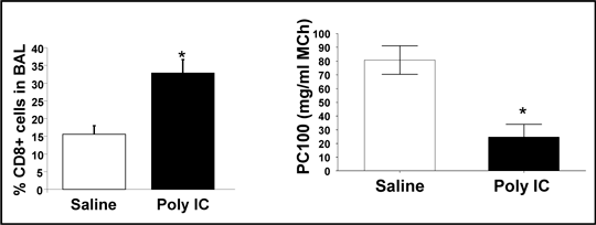
Figure 1. Data showing increase percentage of CD3/CD8+ cells in the BAL fluid of poly I:C treated mice and a reduction in PC100 value (* = P < 0.05).
The major histopathological features of this model included alveolar septa expansion as well as peribronchiolar and interstitial inflammatory cell infiltrate. Oral administration of budesonide had no effect on AHR or neutrophil numbers, however it dose-dependently decreased BALF lymphocyte numbers. Conclusion: Numerous studies have used poly I:C to induce exacerbations in animal models of respiratory diseases; however, for the first time we demonstrate poly I:C alone induces airway hyper-responsiveness and a unique inflammatory phenotype that is only partially sensitive to glucocorticoid treatment.
|
|