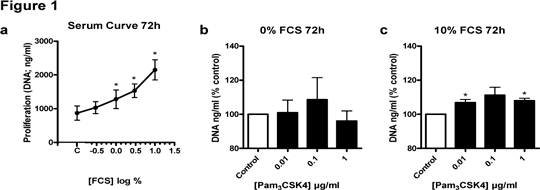
TLR2/1 activation induced proliferation of A549 cells under certain conditions Chronic inflammation and infection are linked with many types of cancer. Toll-like receptors (TLRs) are type I transmembrane receptors that recognize microbial conserved components and initiate an immune response. TLRs promote certain cancer cell proliferation, tumour invasiveness and angiogenesis (1). Here, we investigated whether TLR2 signalling was involved in proliferation of the human lung adenocarcinoma cell line (A549) using the TLR2/1 agonist, Pam3CSK4. Activation of TLRs on A549 cells results in release of the chemokine CXCL8. After 72h treatment, CXCL8 accumulation was measured by ELISA to assess receptor functionality and cell proliferation was determined by measuring DNA content using the CyQuant™ assay. Fetal calf serum (FCS) caused a concentration-dependent increase in A549 cell proliferation after 72h (Figure 1a). Pam3CSK4 (at 1μg/ml) increased CXCL8 release from control levels of 2431 ± 569 pg/ml to 7391 ± 362 pg/ml (72h; n = 6). Pam3CSK4 also caused an increase in proliferation of A549 cells after 72h when media with 10% FCS was used (Figure 1c), but not in media without serum (Figure 1b). Our findings confirm that TLR2 heterodimers expressed on human lung cancer cells are functionally active causing release of CXCL8. In addition, we show that the TLR2/1 agonist Pam3CSK4 also increased/protected cell proliferation. The relevance of this data in terms of lung cancer cell growth remains the subject of our investigation.
Figure 1 
Figure 1. The effects of FCS and Pam3CSK4 on A549 proliferation. A549 cells (in 96 well plates at 104cells per well) were equilibrated for 12 hours in DMEM plus 10% FCS, after which media was replaced as indicated on figure 1 for 72 hours. Cell proliferation was measured using the CyQuantTM assay for quantification of DNA content. Data is mean ± sem of n = 6 replicates measured over 3 separate experimental days. (a) *P<0.05; one-way ANOVA followed by a Dunnett’s multiple comparison post-hoc test. (b) *P<0.05 by a one sample t-test of normalised data.
1 Sato, Y., Goto, Y., Narita, N. and Hoon, D. S. (2009) Cancer Cells Expressing Toll-like Receptors and the Tumor Microenvironment. Cancer Microenviron 2 Suppl 1, 205-214
|
|

