Print version
Search Pub Med
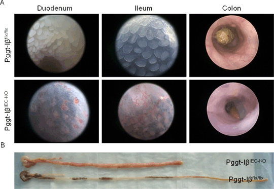
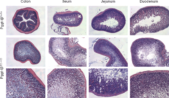
Lack of geranylgeranyltransferase activity in gut epithelial cells leads to disruption of intestinal homeostasis and lethality Introduction: Beneficial therapeutic effects of statins are described in experimental settings of chronic colitis. Regarding the underlying molecular mechanism of statin mediated anti-inflammatory effects, their capacity to interfere with the process of posttranslational protein prenylation seems to be of great importance. Prenylation modifies protein hydrophobicity and affects crucial protein features such as interaction with other proteins or attachment to the cell membrane. Despite the performance of several preclinical studies and clinical trials with specific prenylation inhibitors in cancer, the specific role of prenylation in the pathogenesis of inflammatory bowel diseases has not been defined yet. We decided to analyse the functional in vivo relevance of GGTase-I for intestinal homeostasis and its role within intestinal epithelial cell compartment. Methods: In order to analyse mice with a specific deletion of GGTase-I in intestinal epithelial cells (IEC), floxed Pggt-Iβ mice (Pggt-Iβflx/flx mice, where exon 7 of GGTase-I is LoxP-flanked) were crossed with Villin-Cre mice or Villin-CreERT2 mice in order to generate Pggt-IβIEC-KO mice and its inducible model, respectively. Villin-CreERT2 expression cassette allowed temporally controlled deletion of GGTase-I by injection of tamoxifen. Intestinal phenotype was monitored by endoscopy and histology. Protein expression, cytoskeleton disturbances and caspases activation has been measured by western blot and immunofluorescence stainings. Results: lack of GGTase-I in IEC leads to embryonic lethality before day 14.5 of embryogenesis. Tamoxifen treatment (10 mg/mouse/day during 3 consecutive days) induced the deletion of GGTase-I in IECs of adult mice and successfully reduced the amount of prenylated proteins within IEC, Pggt1bflx/flx Villin-CreERT2 mice developed a severe enteric disease and massive mucosal damage could be visualized by video-endoscopy approximately 7 days after initiation of tamoxifen treatment. Endoscopy and macroscopic analysis show a significant thickening and shortening of the small intestine as well as a proximal-distal gradient, the duodenum being the most affected part of the intestinal tract (Fig. 1). H&E staining indicated a loss of epithelial integrity in the absence of GGTase-1, so that intestinal epithelial cells are not anymore attached to the basal membrane, villi and crypt organisation is disrupted and lamina propria cells are directly exposed to the lumen (Fig. 2). Although Caspase-3 activation can be observed in late time points, a disturbance of cytoskeleton homeostasis cannot be discarded, as it is shown by F-actin staining. Deeper mechanistic studies will show the underlying mechanism. Conclusion: Our results clearly identified a pivotal function of GGTase-I activity in intestinal epithelial cells. Obviously, the posttranslational process of protein prenylation is critically involved in the regulation and maintenance of gut homeostasis. Statin and bisphosphonate effects on epithelial cells may be related to the present findings.
Figure 1: Intestinal phenotype of Pggt1βIEC-KO. (A) Pictures from endoscopy-video of different segments of the gut (B) Macroscopic pictures of isolated gut.
Figure 2: Histological analysis of gut sections from Pggt1βIEC-KO mice. Hematoxylin-Eosin staining.
|


