Print version
Search Pub Med

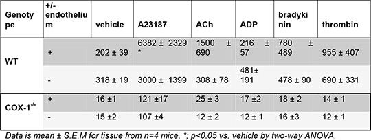
Role of the endothelium and COX-1 in prostacyclin generation by whole vessels stimulated with different agonists Introduction: Prostacyclin is an important cardioprotective hormone produced by the vascular wall, whose synthesis is dependent on cyclo-oxygenase (COX) enzymes. In healthy vessels the endothelium is thought to be the main site of prostacyclin release (Moncada et al 1977). Two isoforms of COX exist, and we have recently published data demonstrating that it is COX-1 rather than COX-2 that drives the production of prostacyclin in mouse aorta (Kirkby et al 2012). In this study we aimed to extend these observations by investigating what proportion of the COX-1 driven aortic prostacyclin production that comes from the endothelium versus the rest of the vessel wall (smooth muscle layers and adventitia). To do this, we explored how removal of the endothelium would influence the ability of aortic tissue to release prostacyclin in response to a range of agonists that are known to activate the endothelium and the vessel wall. Methods: Wild type (WT, C57BL/6 mice) and COX-1-/- mice (n=4, 10-12 weeks old) were killed by CO2 induced asphyxiation. Aortas were removed and divided into 2mm rings after which in some rings the endothelium was removed by rubbing of the luminal surface with forceps. Aortas were allowed to equilibrate in Dulbecco’s modified eagle’s medium (DMEM) for 60 mins at 37°C, before medium was replaced and vessels incubated with A23187 (50µM), bradykinin (100nM), thrombin (1U/ml), ADP (10µM), acetylcholine (ACh, 10µM), or vehicle (0.1% DMSO). After 30 mins, medium was removed for measurement of the stable prostacyclin breakdown product, 6-keto-PGF1 α, by enzyme immunoassay. Results: The release of prostacyclin from COX-1-/- vessels was substantially less (<10%) than that from WT vessels, both under control conditions and in the presence of blood vessel agonists (Table 1). As the levels of prostacyclin production recorded from intact COX-1-/- vessels were towards the limit of detection of our immunoassay it cannot be reliably reported if these were decreased by endothelial cell removal (Table 1). In WT vessels the production of prostacyclin to ACh following removal of the endothelium was reduced by 69±11%; the productions to other agonists were not significantly changed (Table 1). Table 1

Data is mean ± S.E.M for tissue from n=4 mice. *; p<0.05 vs. vehicle by two-way ANOVA. Summary: This data reiterates the overwhelming role of COX-1 in the production of prostacyclin by healthy blood vessels, regardless of the agonist used. It also suggests that the production of prostacyclin in normal vessels is dependent upon both endothelial and non-endothelial cells, but that the relative roles of these sources vary with different agonists. Moncada et al (1977). Thromb Res. Sep;11(3):323-44. Kirkby et al (2012). PNAS In Press.
|