14 day exposure to corticosterone in drinking water results in changes to rat brain mitochondrial function of male wistar rats During periods of chronic stress the organization and regulation of the body’s stress responses is disrupted with elevated levels of glucocorticoids, remaining high [1]. Prolonged exposure of neurons to glucocorticoids reduces their resilience to insults which eventually results in dendritic atrophy in key brain area, fewer neurons and susceptibility to psychiatric disorders [2]. Previous studies in a number of laboratories have shown that both acute and chronic treatment with corticosterone (CORT) impairs oxidative phosphorylation in a number of tissues including rat liver, rat kidneys and in neuronal cell cultures. The present study sought to determine the effect of chronic oral CORT treatment on rat brain mitochondrial function, expressed as respiratory coupling index (RCI). RCI is an indicator of the efficiency of coupling between oxygen consumption (oxidation) and ATP synthesis (phosphorylation), a robust or tight coupling is essential for optimal energy utilization as well as to control or reduce the production of reactive oxygen species (ROS) [3]. Male Wistar rats in the test group were exposed to CORT (50mg L-1) in their drinking water for 14days, while rats in the control group received 0.5% (vv-1) ethanol in their drinking water. On the 14th day animals were sacrificed by rapid decapitation between 9:30 and 10:00 am. Forebrain mitochondria were isolated by differential centrifugation through a percoll density gradient. The RCI was recorded polarographically using a Clarke-type oxygen electrode as described previously in Markham et al., 2012 [4]. Data were analysed using student’s unpaired t-test. 
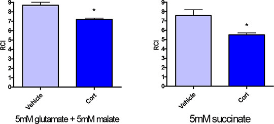
Figure 1 The effect of exposure to corticosterone, on the respiratory coupling index (RCI) of rat brain mitochondria . Complex I substrate 5mM glutamate plus 5mM malate and complex II substrate and 5mM. The bars represent mean ± s.e.m (n = 5). Vehicle=0.5% (vv-1) ethanol, Cort = corticosterone (50mg L-1). *P<0.05 compared to vehicle as control. Fig 1 confirms that chronic exposure to CORT in drinking water significantly (P<0.05; n=5) reduced the RCI of rat brain mitochondrial preparations in relation to both complex I and II In the presence of 5mM glutamate plus 5mM malate the RCI was reduced from 8.70 ± 0.30 to 7.17 ± 0.17; representing a change of 17.6% in complex I supported respiration. Similarly, in the presence of 5mM succinate the RCI was reduced from 7.56 ± 0.60 to 5.50 ± 0.20 (P<0.05; n=5); representing a change of 27.3% in complex II supported respiration. The observed decrease in brain respiratory coupling efficiency is of major significance and clearly indicates that exposure to a glucocorticoid can modify mitochondrial oxygen utilisation and associated intracellular energy production. Such changes in organelle energy efficiency could have profound consequences in terms of neuronal metabolism, survival and plasticity leading to the manifestation of stress related CNS conditions. [1] Manoli, I., etal., (2007). Trends Endocrinol Metab. 18(5), 190-198 [2] Goshen, I., & Yirmiya, R. (2009). Front. Neuroendocrinol. 30, 30-45 [3] Mattson, M.P. (2005). Annu. Rev. Nutr. 25, 237-260 [4] Markham, A., et al., (2012). Eur. J. Neurosci. 35, 366-374
|