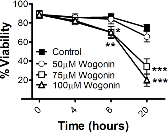
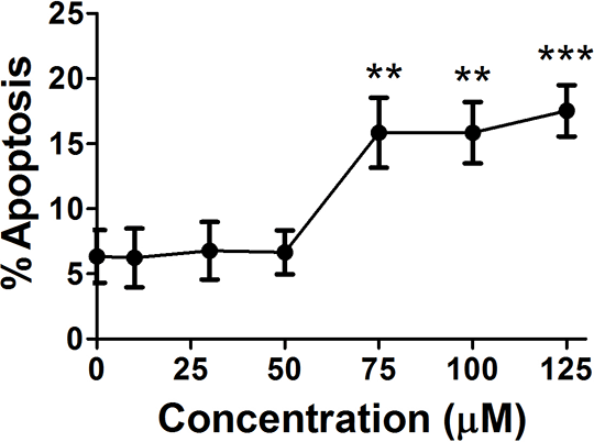
Wogonin induces caspase-dependent human eosinophil apoptosis Introduction: Eosinophils are key effector cells in allergic inflammatory diseases. Their infiltration and accumulation into tissues is implicated in the tissue damage and remodelling seen in diseases such as asthma (1). Glucocorticoids, the mainstay of current treatment for allergic inflammation, cause programmed cell death (apoptosis) of eosinophils. However, their adverse side effects necessitate the need to find alternative treatment options. Here, we investigated whether the flavone wogonin, which has previously been shown to induce neutrophil apoptosis in vitro and in vivo (2), could induce apoptosis of eosinophils in vitro. Methods: Primary human eosinophils were isolated from blood of healthy donors by Percoll-gradient centrifugation and anti-CD16 immunomagnetic separation to remove neutrophils. Eosinophils were cultured with the naturally-occurring polyphenolic flavone wogonin (5,7-Dihydroxy-8-methoxyflavone; 0-125µM); caspase-inhibitors Q-Val-Asp(non-omethylated)-OPh (Q-VD; 20µM) or Z-Val-Ala-DL-Asp(OMe)-fluoromethylketone (zVAD; 100µM); eosinophil pro-survival factors interleukin-5 (IL-5; 0.1ng/mL) or granulocyte-macrophage colony-stimulating factor (GM-CSF; 1ng/mL) before viability and apoptosis assessment were conducted by flow cytometry (Annexin-V/propidium iodide binding) and morphological analysis. Appropriate concentration of drug solvent, dimethyl sulfoxide (DMSO), was added to controls. Data (mean ± SEM) were analysed by 1-way ANOVA with a Dunnett’s test, or t test as appropriate. Results: Wogonin induced time- and concentration-dependent death of primary human eosinophils (6h control viability 86.1±4.2% vs. 70.2±6.4% 75µM wogonin; p<0.05) (Figure 1). This was secondary to wogonin-induced eosinophil apoptosis, observed at 6 hours (Figure 2). This gave an EC50 of 63.4µM wogonin at 6h and 62.8µM wogonin at 20h. Wogonin-induced eosinophil apoptosis was dependent upon activation of caspase enzymes as co-incubation with caspase inhibitors (z-VAD and Q-VD) prevented wogonin-induced apoptosis (6h 75µM wogonin apoptosis 15.2±2.2% vs 6.1±1.1% and 7.9±3.6% Q-VD+75µM wogonin & zVAD+75µM wogonin, respectively, p<0.05). Wogonin was still able to induce eosinophil apoptosis even in the presence of the pro-survival factors IL-5 and GM-CSF (p<0.05). These data demonstrate that wogonin induces eosinophil apoptosis, and therefore has potential for promoting resolution of eosinophillic inflammation in human allergic disease. *p<0.05, **p<0.01, ***p<0.001 
Figure 1 
Figure 2 (1) Leitch AE et al, Mucosal Immunol 1:350, 2008, (2) Lucas CD et al, FASEB J 27:1084, 2013
|


