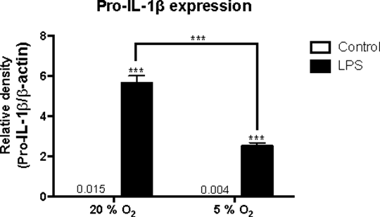
Lipopolysaccharide regulation of the NLRP3 inflammasome and proIL-1ββ under normoxic versus hypoxic conditions in cultured (BV-2) mouse microglia Introduction: The NLRP3 inflammasome is a multi-protein complex responsible for caspase-1 activation leading to cleavage of proIL-1 and proIL-18 (1). Inflammation has been implicated in neurological disorders, such as Alzheimer’s disease, where microglia activation and enhanced NLRP3 inflammasome signalling are evident (2). In this study, expression of NLRP3 inflammasome signalling following Toll-like receptor 4 (TLR4) activation by lipopolysaccharide (LPS) was assessed in cultured mouse microglia. While most in vitro studies use 20 % O2, we have compared this with 5 % O2 to more accurately mimic oxygen availability in the brain, which can range from 0.5 - 7 % (3). This study aimed to compare TLR4-dependent expression of NLRP3 and proIL-1β in BV-2 microglia under reduced oxygen conditions (5 % O2) and normal culture conditions (20 % O2). Methods: A murine microglial BV-2 cell line was incubated under 5 % CO2 & air (20 % O2) or 5 % O2, 5 % CO2 and 90 % N2 in the presence or absence of 0.1 µg/ml lipopolysaccharide (LPS) for 4 hours at 37 ˚C. Cell lysates were collected, total protein concentration measured and NLRP3 component expression determined by western blot. Quantification was performed using densitometry measurements and levels normalized to β-actin loading controls. Data analysed by ANOVA with posthoc Tukey’s test. Results: BV-2 cells express a basal level of NLRP3 but not of proIL-1β.. LPS (0.1 µg/ml) increased NLRP3 protein expression (532 ± 8 %) and induced proIL-1β protein expression, compared to vehicle-treated control, under normal culture conditions (20 % O2) (p<0.001, n=3). However, under 5 % O2 culture conditions, the effects of LPS were significantly attenuated; reducing LPS-induced expression of NLRP3 by 23 ± 7 % (p<0.05) and proIL-1β by 55 % ± 5% (p<0.001), compared to vehicle-treated control (n=3). Baseline levels of protein expression under 5 % O2 culture conditions remained unchanged. 
Conclusion: LPS induced the expression of inflammatory proteins involved in NLRP3 inflammasome signalling in BV-2 microglia. When incubated in low oxygen conditions, thought to be similar to the in vivo environment (5% O2), cytosolic NLRP3 and proIL-1β expression was reduced, indicating oxygen sensitivity. This observation could be a result of enhanced processing and secretion of proteins via NLRP3 inflammasome activation. (1) Latz, E. et al. (2013) Nat Rev Immunol. 13(6), 397-411 (2) Heneka, M. T. et al. (2013) Nature. 493(7434), 674-678. (3) Ivanovic, Z. (2009) J Cell Physiol, 219(2), 271-275. Supported by a MRC CASE PhD studentship with Janssen Research and Development, a Division of Janssen Pharmaceutica NV
|