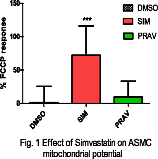
Role of mitochondria in the relaxation to simvastatin in porcine isolated coronary artery. HMG CoA reductase inhibitors, or statins, are widely used as cholesterol-lowering agents in the treatment of dyslipidemias. Studies have shown that simvastatin (10µM) have vasorelaxant pleiotropic effects independent of its effects on cholesterol synthesis whereas pravastatin has no effects on vascular tone (Lorkowska et al, 2005, Nagaoka et al, 2007). Simvastatin has been also shown to cause mitochondrial depolarization in skeletal muscle (Sirvent. et al, 2005) and hepatocytes (Abdoliet et al, 2013). Mitochondrial depolarization has been purported to regulate vascular smooth muscle tone via mitochondrial reactive oxygen species-dependent and independent mechanisms (Katakam et al, 2013). Therefore, the present work aimed to determine the direct effects of simvastatin on the isolated porcine coronary artery (PCA) and to identify the acute effect of statin on mitochondrial membrane potential (Δψm) in human aortic smooth muscle cells (ASMC). Coronary arteries from pig hearts obtained from a local abattoir were mounted in isolated tissue baths and contracted with U46619, a thromboxane A2 agonist prior to addition of a single concentration of simvastatin or pravastatin (10µM) in the absence or presence of the mitochondrial complex I inhibitor rotenone (10µM) and the complex III inhibitor myxothiazol (10µM). Human ASMC (Lonza) were grown on sterilized glass cover slips, between passages 7 and 9, loaded with 10µg ml-1 rhodamine123 (Rh123) at 37 °C for 10 min in Hanks balanced salt solution containing 5 mM glucose (pH 7.4) to achieve a quenching concentration of the dye within the mitochondrial matrix. Cover slips were washed and placed in a superfusion system. Changes in fluorescence of Rh123 (excitation 480nm, emission 530nm) during perfusion with (10µM) simvastatin or pravastatin was measured at 32 °C for about 10min. Control cells were perfused with vehicle only (0.1% DMSO). 
Simvastatin 10µM produced a time-dependent relaxation of the porcine coronary artery 75.4± 4.4% relaxation at 120 mins, mean ± SEM from 9 different animals; P <0.001 (2- way ANOVA followed by Bonferroni post-hoc test), which was inhibited by a combination of rotenone 10µM and myxothiazol 10µM (p<0.005, 2-way ANOVA). In ASMC grown in culture, simvastatin 10µM produced an increase in Rh123 fluorescence, n=15 separate experiments, (one-way ANOVA using Tukey\\\'s multiple comparisons test), which is indicative of a change in Δψm (fig. 1). By contrast neither pravastatin nor vehicle (0.1% DMSO) had any significant effect on the coronary tone or the smooth muscle mitochondrial potential. These data demonstrate that the lipophilic, simvastatin, but not the hydrophilic, pravastatin, produces a relaxation of the porcine coronary artery. Inhibition of the relaxation with mitochondrial complex inhibitors suggests that the mitochondria are involved in this response. The data showing changes in Δψm in cultured smooth muscle cells provide further evidence that simvastatin can act on mitochondria in vascular smooth muscle. As pravastatin had no effect, this may be related to the lipophilicity of the compounds. Abdoliet et al., (2013) J Biochem Mol Toxicol. 27:287–294 Katakam et al., 2013 Arterioscler Thromb Vasc Biol. 33:752-759 Lorkowska et al., (2005) Prostag Leukot Essent Fatty Acids. 72:133-8 Nagaoka et al., (2007) Invest Ophthalmol Vis Sci. 48:825-32. Sirvent.et al., (2005) Biochem. Biophys. Res. Commun. 329:1067–1075
|