Print version
Search Pub Med

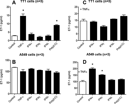
Modulation of endothelin-1 release by TNFα and interferons from human lung alveolar type I versus alveolar type II epithelial cells Endothelin (ET)-1 is a vasoconstrictor, bronchoconstrictor and mitogen that is associated with pulmonary hypertension(Shao et al., 2011), asthma and COPD(Polikepahad et al., 2006). Under control conditions ET-1 is released by endothelial cells in relatively high amounts (≈100pg/ml) but in low levels from most other cell types. Our group was the first to describe ET-1 as an interferon (IFN) inducible gene(George et al., 2012a). IFNs are critical mediators of the innate immune response to viruses. Thus, we have suggested that viral infection, via IFN release, can result in ET-1 mediated inflammation in the vasculature and lungs(George et al., 2012a). The relative ability of lung epithelial cells to release ET-1 is not well studied, but clearly relevant to our understanding of the role of this hormone in the lungs. The purpose of this study was to investigate the effects of types I, II and III IFN on ET-1 release by two human lung epithelial cell lines; A549 cells (an adenocarcinoma cell line with type II pneumocyte characteristics) and TT1 cells, a recently created immortalised type I pneumocyte cell line(Thorley et al., 2011). Responses were compared with those induced by POLY(I:C), which activates viral Toll like receptor 3 and performed with or without TNFα, which primes IFN responses in some cells(George et al., 2012b). Cells were cultured as described previously(Thorley et al., 2011) and stimulated with TNFα(10ng/ml), type I IFNα(10ng/ml), type II IFNγ(10ng/ml), type III IFNλ(1µg/ml) or POLY(I:C)(100µg/ml) for 24hs(Fig 1A & B). In separate experiments, cells were co-stimulated with TNFα (Fig 1C & D) and IFNs or POLY(I:C). ET-1 was measured by ELISA. Figure 1: ET-1 release from TT1 (A, C) or A549 (B, D) stimulated for 24 hours. Data is mean ± SEM; n=3. *p<0.05 by one-way ANOVA and Dunnett post-test. Basal ET-1 release was low from TT1 cells, but unexpectedly high from A549 cells. ET-1 release was increased from TT1, but not A549 cells by TNFα and POLY(I:C). In the presence of TNFα, IFNα and IFNγ, but not IFNλ or POLY(I:C) induced ET-1 release from A549, but not TT1 cells. These data reveal lung alveoli epithelium as a rich source of cytokine/IFN-induced ET-1 and have relevance to our understanding of sites of production outside the vasculature in IFN driven inflammation. George PM, Badiger R, Alazawi W, Foster GR, Mitchell JA (2012a). Pharmacol Ther 135(1): 44-53. George PM, Badiger R, et al. (2012b).. Biochem Biophys Res Commun 426(4): 486-491. Polikepahad S, Moore RM, Venugopal CS (2006). Res Commun Mol Pathol Pharmacol 119(1-6): 3-51. Shao D, Park JE, Wort SJ (2011). Pharmacol Res 63(6): 504-511. Thorley AJ, Grandolfo D, Lim E, Goldstraw P, Young A, Tetley TD (2011). PLoS One 6(7): e21827. |