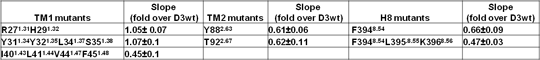
The quaternary structure of dopamine D2 and D3 receptors: comparisons of the homo-oligomerisation of two closely related G protein-coupled receptors The dopamine receptors, D2-long isoform (D2L) and D3, are rhodopsin-family GPCRs. Disruption of dopaminergic neuro-transmission is implicated in multiple disorders including Parkinson’s disease and schizophrenia but, because of the overlap of ligand recognition and co-expression of the two receptors, their individual contributions are challenging to define; moreover, the capability of both D2L and D3 to form homo- and hetero-oligomers can also influence dopaminergic neuro-transmission. While the capacity of the D2L receptor to form homo-dimers and higher-order oligomers has been studied extensively (1, 2), less is known about D3-homo-interactions or D2L-D3 hetero-interactions (2), Homo-oligomerisation of the D3 receptor was investigated to examine similarities and differences with D2L. Initially, Homogenous Time Resolved-FRET (htrFRET) using Tag-lite technology (3) was employed. In cells expressing SNAP-D3 receptor, combinations of SNAP-Lumi4-Tb (10 nM) as energy donor and increasing concentrations of the corresponding htrFRET energy acceptor SNAP-Red, followed by excitation at 337nm, resulted in fluorescence output at 665nm as a consequence of energy transfer between SNAP-Red and SNAP-Lumi4-Tb bound to the SNAP tagged receptor. The resulting fluorescent signal at 665nm reflects the close proximity between D3 receptors and their likely direct physical interaction. Alanine scanning mutagenesis combined with htrFRET was then employed to investigate the roles of residues in transmembrane domain (TM) 1, TM2 and the intracellular Helix 8 (H8) of the D3 receptor in the formation of at least one interface of D3 receptor homo-oligomers. Wild type D3 and D3 receptor mutants, modified at the N-terminus with SNAP-tag, were expressed transiently in HEK293T cells. Cells expressing different levels of the receptor variant of interest were grown on 96 well plates and labelled with SNAP-Lumi4 and SNAP-Red. Fluorescence emission at 620 nm (indicative of cell surface expression of the receptor construct) and 665nm (reflecting protein-protein interaction) was then measured concurrently and correlated. For wild type and each mutant studied this resulted in a linear relationship. However, the slope of the linear regression for a number of the mutants was reduced substantially compared to that for the wild type receptor (Table), indicative of reduced proximity between D3 receptor monomers and hence alteration of receptor oligomer structure.

Residues altered to Alanine are identified by the ‘one-letter’ code and by both position in the primary amino acid sequence of the human D3 receptor and the corresponding Ballesteros and Weinstein residue location numbering system. Data are given as the mean ± S.E. of 3 independent experiments. These results were then utilised to generate a molecular model showing the D3 receptor to form protein-protein interactions via an interface consisting of H8-H8, TM1-TM1 and TM2-TM2 interactions that are not conserved in D2L receptor homo-dimers. These studies will facilitate the identification of strategies and, potentially, small molecule ligands able to disrupt either homomers or heteromers involving the D3 receptor to assess and understanding specific physiological roles of GPCR homomers and heteromers. (1) Guo W et al, EMBO J 27:2293, 2008 (2) Pou C et al, J Biol Chem 287:8864, 2012 (3) Maurel D et al, Nat Methods 5:567, 2008
|