Novel histamine H1-receptor fluorescent antagonists for the study of receptor-ligand interactions G protein-coupled receptor drug discovery focuses on characterising the affinity and efficacy of a compound at a given receptor but rarely takes into account the kinetics of ligand association and dissociation. There is growing evidence to suggest that the kinetics of ligand binding can be very important in vivo (Vauquelin and Charlton, 2010). It has recently been shown that the dissociation kinetics of a fluoresecent GPCR agonist can be directly monitored using confocal microscopy coupled to a perfusion system which allows constant flow of buffer over the cells to prevent rebinding of the ligand (May et al). Here, we developed a range of fluorescent histamine H1 receptor (H1R) antagonist, based on two different parent pharmcophores and characterised their pharmacology and dissociation rates. Eight fluorescent fluorescent histamine antagonists were synthesised in this study, five based on the histamine H1 antagonist mepryamine and three on the recently described antagonist VUF131816 (de Graaf et al, 2011). These were coupled to the BODIPY-630/650 or BODIPY-FL fluorophores via a peptide based linker and purified (to >99%) by RP-HPLC. Affinities were determined as follows; CHO cells expressing the H1R were incubated with a fixed concentration of fluorescent compound for 45 min prior to addition of increasing concentrations of histamine and the change in calcium monitored and pKB values calculated from the resulting shift in the histamine concentration response curve using the Gaddum equation and EC25 values. Dissociation rates were determined using a Zeiss 710 confocal microscope coupled to a perfusion system as described in May et al. (2010) and experiments were performed for 5 minutes at 37oC with a flow rate of 7 ml per min.

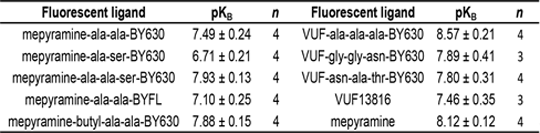
Table 1 Affinity values of fluorescent H1 antagonists and parent pharmacophores used in this study As shown in Table 1, all the fluorescent ligands tested retained affinity at the H1R compared to the parent pharmacophores. Confocal imaging showed that displaceable membrane binding, with low levels of non-specific intracellular accumulation of the ligands could be detected for all of the eight fluorescent ligands. Kinetic experiments indicated that the fluorescent mepryamine compounds have a very slow dissociation rates at the H1R receptor and over the time course of the experiments (5 min) very little of the VUF based compounds have dissociated from the H1R. These data indicate that ligands with similar affinity display different dissociation kinetics at the H1R. Vauquelin G & Charlton S, Br J Pharmacol 161:488, 2010 de Graaf et al,J Med Chem 54: 8195, 2011 May et al, Mol Pharmacol 78: 511, 2010 This work has received support from the EU/EFPIA Innovative Medicines Initiative Joint Undertaking, K4DD grant n° 115366.
|