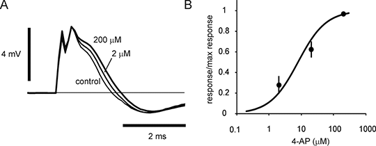
S-fibres in ex vivo rat optic nerve are ultra-sensitive to two K+ channel blockers 4-aminopyridine (4-AP; ampyra,dalfampridine) is useful in the treatment of walking impairment in multiple sclerosis (MS) (1). However, the classically hypothesised mechanism of action, that of widening action potentials in axons with paranodal or segmental demyelination, is not convincing (2). This is because the useful concentration range of drug in vivo is too low to block the voltage-gated K+ channels involved (3). More recent experiments on central axons have revealed fibre sub-types with differing pharmacology, including cerebellar parallel fibres exhibiting a resting sensitivity to 4-AP sufficiently low to be affected by the drug at clinically useful concentrations (4). Optic nerve contains a sub-set of slowly conducting axons (S-fibres) that are very sensitive to 4-AP. This sensitivity reveals itself in an increased response to brief applied stimulus currents (e.g. Fig 1), and indicates that the resting properties of the normal axons are changing on exposure to low concentrations of the K+ channel blocker.
Figure 1. A, Low concentrations of 4-AP act on the late component of normal compound action potentials recorded from ex vivo rat optic nerve, B, data plotted as means ± sem (n = 4 nerves). Smooth curve is a rectangular hyperbola plotted with a Kd of 8 μM. The action is attributable to an increase in S-fibre response to brief applied stimuli (200 μs). Similar findings were obtained with tetraethylammonium (TEA) ions. Kd (µM; estimated in individual nerves) for 4-AP = 11.29 ± 3.58, TEA = 10.52 ± 2.64; mean ± sem, n = 4,4, respectively. Optic nerves were isolated from Wistar rats (300 - 400 g), mounted in an ex-vivo bath, and stimulated using constant-current pulses applied via a suction electrode. Antidromic compound action potentials were recorded across a Vaseline gap. The rheobase of the S-fibres is long (~3 ms at 30 °C), and similar fibres are found elsewhere in the CNS, including the spinal cord (5). The data are interpreted as suggesting that blocking K+ channels reduces rheobase duration, and enhances S-fibre recruitment to a brief stimulus, possibly by depolarizing the axons through a block of K+ channels open at rest. Such an action could conceivably aid action potential invasion of pre-synaptic terminals. (1) Hadavi, S et al MSARD 3: 17, 2013 (2) Bostock, H et al J Phyiol 313: 301, 1981 (3) Smith, KJ et al Brain 123: 171, 2000 (4) Alviña, K and Khodakhah, K J Neurosci 30: 7258, 2010 (5) West, DC and Wolstencroft, JH J Physiol 337: 37, 1983
|