Print version
Search Pub Med
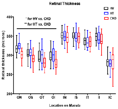
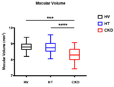
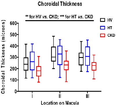
Clinical Utility of Optical Coherence Tomography in Patients at high CVD risk Background: Compared to the general population, patients with chronic kidney disease (CKD) have a 10-30 fold increased risk of mortality due to cardiovascular disease (CVD). There is an established association between the vasculopathy affecting the kidney and the retina, suggesting common pathological mechanisms. Even after correcting for renal dysfunction and classical CVD risk factors, retinopathy is associated with a higher prevalence of CVD. Optical coherence tomography (OCT) is a novel, non-invasive and rapid method of cross-sectionally imaging the retinal and choroidal structures. Its use in patients at high CVD risk has not been previously explored. Methods: We used the new SPECTRALIS OCT to perform an exploratory study examining the retinal and choroidal structures in 24 patients with hypertension, 24 with varying degrees of CKD and 25 age and sex matched healthy controls. The same ophthalmologist carried out a single OCT scan (comprising of 3 images) in each subject. Measurements included retinal thickness, retinal nerve fibre layer (RNFL) thickness, macular volume and choroidal thickness. Retinal thickness was automatically calculated in 9 separate locations on the macula whereas choroidal thickness was manually calculated in three separate locations on the macula. Results: Retinal thickness was reduced across the outer 4 locations on the macula in CKD patients compared to both those with hypertension (p<0.01), and healthy volunteers (p<0.05) (Figure 1). RNFL thickness did not differ between groups. Macular volume was lower in CKD patients compared to both patients with hypertension (p<0.0001) and healthy volunteers (p<0.001) (Figure 2). Similarly, CKD was associated with a reduced choroidal thickness (across all three separate locations on the macula, Figure 3) compared to both patients with hypertension (p<0.001) and healthy volunteers (p<0.01). Interestingly, in those with CKD, a thinner choroid was associated with a lower glomerular filtration rate (p< 0.01) and more severe proteinuria (p<0.01), both important independent CVD risk factors.
Figure 1
Figure 2
Figure 3 HV: healthy volunteer; HT: hypertension; CKD: chronic kidney disease Conclusions: The decreases in retinal and choroidal thickness seen in CKD may represent significant systemic microvascular injury. It remains unclear how these may change with therapy. Thus, OCT may offer potential utility in assessing microvascular damage in patients at high CVD risk as well as a potential biomarker of efficacy for therapies used to reduce this risk.
|


