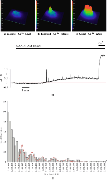
Glucose and NAADP trigger Elementary Intracellular Ca 2+ Signals in Pancreatic β -Cells Figure 1 Intensity profiles of a β-cell cluster (a) at baseline, (b) in the presence of an elementary calcium event, (c) during global calcium influx. (d) Representative trace of a β-cell showing calcium events triggered by NAADP-AM. Maximum intensity change after normalising to baseline is plotted against time. Red line denotes baseline average. Extracellular calcium was re-admitted at the end of the experiment; leading to a global calcium response. (e) Quantal analysis of elementary calcium events. Frequency histogram of event amplitude across areas of interest within a single representative cell (n=51). Note: Histogram was cropped for better resolution (maximum intensity bin is 2.09-2.095). The peaks of the putative modal distribution were selected to fit a quintuple poly-Gaussian function (sum of five Gaussian functions with means, SDs: 0.143, 0.007; 0.171, 0.008; 0.201, 0.0089; 0.235, 0.0092; 0.270, 0.0103, respectively). The putative unitary event amplitude thus lies at ∼0.03 ΔF/F0.
(1) Ashcroft FM et al. (1984). Nature 312: 446–448. (2) Axelrod D (1981). J Cell Biol 89: 141–145. (3) Cheng H et al. (1993). Science 262: 740–744. (4) Cook DL and Hales CN (1984). Nature 311: 271–273. (5) Henquin JC (1998). Endocrinology 139: 993–998. (6) Johnson JD and Misler S (2002). Proc Natl Acad Sci USA 99: 14566–14571. (7) Masgrau R et al. (2003). Curr Biol 13: 247–R251. (8) Parker I and Ivorra I (1990). Science 250: 977–979. (9) Parker I et al. (1996). Cell Calcium 20: 105–121. (10) Seghers V et al. (2000). J Biol Chem 275: 9270–9277.
|