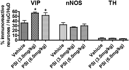
Increased VIP expression in rat jejunum following systemic administration of proteasomal inhibitor I (PSI). Introduction:Gastrointestinal (GI) disturbances, including reduced motility, gastroparesis and constipation, are an early sign of Parkinson’s disease (PD) (1, 2). They can be difficult to treat and adversely affect the absorption of dopaminergic medication. Toxin-induced animal models of PD are widely used to study the motor dysfunction but there has been little investigation into changes in GI function. The proteasomal inhibitor, PSI mimics many of the central pathological changes of PD after its systemic administration (3), but whether alterations occur in the GI tract is not known. Vasoactive intestinal peptide (VIP) and nitric oxide are key neurotransmitters regulating peristalsis and a change in their relative activities can alter GI motility. For this reason, the present study investigates the effects of PSI administration on the number of VIP+ve and neuronal nitric oxide synthase (nNOS)+ve neurons in the myenteric plexus of the jejunum in the rat. Methods: Female Wistar rats (200-220g, N=6; Harlan, UK) were administered either PSI (3.0 or 6.0 mg/kg, sc; (4)) or its vehicle (100% DMSO; Sigma-Aldrich, UK) once a day on days 1, 3, 5, 8, 10 and 12. On day 34, animals were overdosed with sodium pentobarbital and perfused with 0.1M phosphate buffered saline (pH7.4). Brain and jejunum were dissected and post-fixed in 4% paraformaldehyde (pH7.4) at 4°C. Brain tissue was cryoprotected in 30% sucrose and cut into free-floating sections (30 µm) and TH+ve cell number was determined in the substantia nigra pars compacta (SNpc) using immunohistochemistry. Jejunum was processed into paraffin-embedded tissue blocks and cut into sections (6.0 µm) and stained with antibodies against tyrosine hydroxylase (TH), HuC/D, nNOS and VIP using immunohistochemistry. Data are presented as mean ± SEM and analysed by one-way ANOVA and Newman Keuls multiple comparison test. Results: In SNpc the number of TH+ve neu
Conclusion: Inhibition of the proteasome following PSI decreased in the number of dopaminergic cell bodies in the SNpc, as expected, and increased the number of VIP+ve neurons in the myenteric plexus of the jejunum. Increased VIP+ve and reduced nNOS+ve neurons have also been reported in the ileum and colon of 6-OHDA-lesioned animals (5), suggesting the neurochemical plasticity may be centrally mediated. However, the role of nNOS+ve remains to be fully elucidated. Further investigations are required to determine the physiological effect of increased VIP activity in the PSI model of PD. (1) Mollenhauer et al. (2013) Neurology 81: 1226-1234 (2) Del Tredici and Braak (2012) Mov Dis. 27: 597-607 (3) McNaught et al. (2004) Ann Neurol 56: 149-162 (4) Soltani et al. (2014) Bioorg Med Chem in press (5) Colucci et al. (2012) Auton Neurosci. 169: 77-86.
|