Print version
Search Pub Med

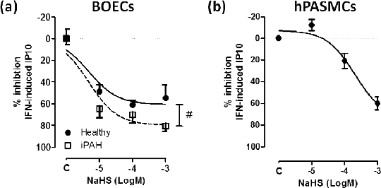
Hydrogen sulphide inhibits IP10 release from interferon stimulated endothelial cells and pulmonary vascular smooth muscle cells: relevance to pulmonary arterial hypertension Pulmonary arterial hypertension (PAH) is a rare but severe disease with limited treatment options. The role of vascular inflammation in PAH is increasingly recognized. We have previously shown that interferon-α (IFN-α) and IFN-induced protein 10 (IP10) correlate with biomarkers and disease indicators in PAH1. We have also shown that IFN-α activates IP10 release from endothelial cells grown out from blood progenitors (blood outgrowth endothelial cells, BOECs) from donors with PAH1 as well as from human pulmonary artery smooth muscle cells (hPASMCs)2. IP10 is increasingly recognised as an inflammatory mediator and a potential therapeutic target in the treatment of PAH3. Hydrogen sulphide (H2S) is an endogenously produced vasoactive gasotransmitter with anti-inflammatory properties that has recently been suggested as a potential therapy for PAH4. Here we have explored the effects of H2S on IP10 and ET-1 (an established therapeutic target in PAH and an IFN-induced gene3) release from BOECs of healthy donors and patients with idiopathic PAH (iPAH) and from hPASMCs. BOECs were isolated and cultured as previously described. hPASMCs were isolated from explants of histologically normal lung tissue obtained from patients without PAH, undergoing lobectomy, as described previously2. IP10 and ET-1 levels were measured by ELISA. As we have shown before, IFN-α (10 ng/ml; 24 hours) increased IP10 (pg/ml) release from BOECs1 healthy donors (basal 31.8±10.0; IFN-α 557.5±74.8) or from patients with iPAH (basal 30.1±10.7; IFN-α 994.6±459.2) and co-released IP10 (basal 40.6±8.4; IFN-α 577.5±205.8) plus ET-1 from hPASMCs2 (p<0.05, paired t-test, n=6). Co-incubation of cells for 24 hours with the H2S donor NaHS (10-7-10-3 M) in the presence of IFN-α (10 ng/ml) caused a concentration-dependent inhibition of IP10 release from all both types tested (Figure 1). NaHS was more potent as an inhibitor of IFN-α induced IP10 from BOECs from patients with iPAH. NaHS had no effect on ET-1 release from either cell type under any condition tested (p>0.05, one-way ANOVA; n=6).
Figure 1: Effect of H2S on IFN-α induced IP10 release by (a) blood outgrowth endothelial cells (BOECs) cultured from healthy donors or from patients with idiopathic pulmonary hypertension (iPAH) and (b) human pulmonary artery smooth muscle cells (hPASMCs). Data is mean ± SEM for n=6. #p<0.05 two-way ANOVA. This data supports the idea that H2S may be protective and a therapeutic option in PAH. (1) George PM et al. (2014) Circ Res 114: 677-688 (2) Badiger R et al. (2012) PLoS One 7: e46779 (3) George PM et al. (2012) Pharmacol Ther 135: 44-53 (4) Ariyaratnam P et al. (2013) Microvasc Res 90: 135-137 (5) Starke RD et al. (2013) Blood 121: 2773-2784.
|