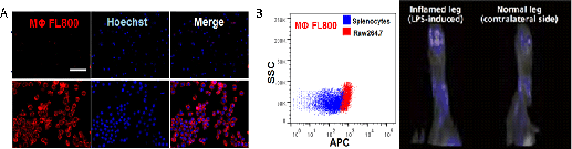
Non-Invasive Imaging of Acute Inflammation by Multispectral Optoacoustic Tomography (MSOT) Macrophages play important roles in development as well in adaptive and innate immune responses. Infiltration and accumulation of macrophages are processes observed in various disease states, including infection, injury, tumorigenesis, and cardiovascular and metabolic diseases. In vivo imaging of macrophage distribution and accumulation in inflamed sites is therefore a powerful tool for enabling diagnosis as well as monitoring therapy response. The aim of the work described here, was to develop a molecular imaging agent for characterizing macrophage-infiltration in LPS-induced inflammation by Multispectral Optoacoustic Tomography (MSOT). MSOT imaging is based on the generation of ultrasound waves induced by the absorption of light pulses by tissue. During MSOT imaging, an object is illuminated using a near-infrared (NIR) laser at multiple wavelengths (range: 680 – 980nm) to then spectrally resolve distinct absorbers. Absorption by intrinsic tissue chromophores (e.g. oxygenated and deoxygenated hemoglobin, melanin) provides rich anatomical contrast, while signal from exogenously administered contrast agents can be multi-spectrally resolved simultaneously allowing for functional and molecular imaging (1). For this study, acute inflammation was induced in adult male C57BL/6J mice, by injecting LPS in one of the hind paws, using the contralateral side as a control. Macrophage-rich regions in the hind legs of mice were then visualized by a newly developed molecular imaging agent, xPLORE MΦ FL800, which was identified through screening a diverse library of NIR ligands for binding specifically to macrophages vs. undifferentiated splenocytes. MSOT imaging was used to evaluate the pharmacokinetics and biodistribution behaviour of xPLORE MΦ FL800. The molecular imaging agent displays fast clearance from the blood circulation (t1/2 = 21mins) with retention at macrophage-rich regions, enabling short imaging sessions. In the LPS-induced model of acute inflammation, increased retention of the probe in the inflamed leg vs. the contralateral non-LPS-treated leg was observed as early as 20 mins. post injection (0.1 ± 0.1 MSOT Units vs 0.5 ± 0.1 MSOT Units) and contrast enhancement was retained for up to 5 hrs. In summary, MSOT offers a new and unique imaging modality that (1) has a resolution at least ten-fold higher than nuclear and optical imaging, (2) allows for real-time imaging with molecular specificity through several centimetres of tissue and (3) is safe (i.e. no ionizing radiation) and cost-efficient. We used this modality, in combination with xPLORE MΦ FL800, to detect inflammatory sites characterized by macrophage infiltration in vivo.
(1) Razansky D et al. (2011). Nat. Protoc 6:1121-9. (2) Kang NY et al. (2014). Chem. Comm 50:6589-91.
|