THE INFLUENCE OF DOXORUBICIN AND PACLITAXEL ON THE ACTIVITY AND EXPRESSION OF CYTOCHROME P450s IN MOUSE LIVER MICROSOME
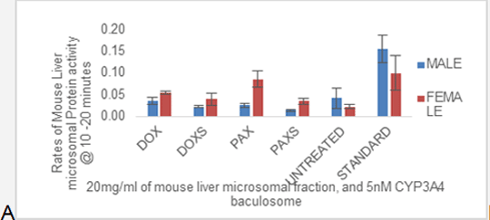
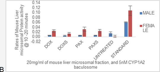
Proteins are primary effectors of drug action; hence it is essential to study how drugs can affect protein expression, and activity (1). In cancer research, it is common to study the effects of anticancer drugs on proteins and pharmacoproteomics plays an important role in determining the effects of anti-cancer drugs on protein expression. More so, the effect of drugs on protein activity can be studied using enzymatic assays. Previous research found doxorubicin to inhibit both CYP1A2 and CYP3A4 activity (2). However, another research showed that doxorubicin induces CYP1A2 activity (3). These differences may be due to variance in species. On the other hand, paclitaxel was found to be a potent inducer of CYP3A in rats (4). Based on these findings one can speculate that doxorubicin may inhibit CYP3A4, and induce CYP1A2, whereas, paclitaxel may induce CYP3A4 homolog in mice. Male and Female H: NU nude mice (n=30 including untreated mice) were treated peritoneally for 24 hours with 20mg/kg paclitaxel, paclitaxel solvent (10% DMSO, 10% Cremophor oil, and 80% saline), and intravenously with 10mg/kg doxorubicin, and doxorubicin solvent (saline) by Home Office licensed staff at the Institute of Cancer Therapeutics, University of Bradford. The influence of doxorubicin, and paclitaxel administration on the activities of CYP1A2 and CYP3A present in mouse liver microsomes was analysed using vivid assays. Given that vivid assays are designed for human CYP1A2 and CYP3A4, the ability to use vivid assays for mouse enzymes was novel. Vivid assay revealed that female mice had generally higher activity than male mice. The exact subtype of CYP affected could not be determined due to the lack of specificity of the vivid substrates. However, the protein activity observed was evidently due to the presence of CYP450 enzymes in the samples. There was a significant difference between treated (0.01/min ± 0.00 – 0.03/min ± 0.01) and untreated (0/min ± 0.00) female samples (p ≥ 0.05) analysed with CYP1A2 vivid substrate (EOMCC). However, treated (0.01/min ± 0.00) and untreated (0.01/min ± 0.01) male samples showed no significant difference. The results were similar after analysis with CYP3A4 vivid substrate (BOMCC). The values represent mean ± SEM, and significance was tested using ANOVA analysis.
In conclusion, it is possible to use mouse models for vivid assays although some improvements may be necessary. Moreover, CYP450s seemed to have been induced by doxorubicin and paclitaxel in female samples, due to higher activity rates in treated samples when compared with untreated samples. ABBREVIATIONS: CYP1A2, CYP3A4, CYP3A11, CYP3A – CYTOCHROME P450 (CYP), Family (1, 3), Subfamily (A), Polypeptide (2, 4, 11); DOX – Doxorubicin; PAX – Paclitaxel; DOXS – Doxorubicin solvent; PAXS – Paclitaxel solvent; EOMCC - ethoxymethyloxy-3-cyanocoumarin; BOMCC - 7-benzyloxymethyloxy-3-cyanocoumarin REFERENCES: (1) Twyman (2004), Principles Of Proteomics. New York: BIOS Scientific Publishers, 2004. (2) Zordoky and Elkadi (2008). Vascular Pharmacology 2008, 49 (4-6), 166-172. (3) Masek et al. (2011). Drug Metabolism and Disposition 2011, 39 (9), 1704-1710. (4) Zordoky et al. (2011). Drug Metabolism And Disposition 39: 1440-1450.
|