Catecholamines Facilitate Cardiac Arrhythmias During Low Flow Global Ischaemia In Rat In Vitro
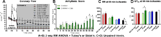
In the rat Langendorff perfused heart, which is nominally denervated, perfusion with added catecholamines facilitates regional ischaemia-induced ventricular arrhythmias when baseline arrhythmia severity is low due to small ischaemic zone (IZ) size (1). We have now tested whether arrhythmia facilitation occurs when baseline severity is low for a different reason, namely collateral flow (simulated here by low flow global ischaemia) and when catecholamines are delivered discretely to the ischaemic region (achievable with low flow ischaemia). Male Wistar rat (280-360g) hearts were perfused with modified Krebs’ solution (3mM K+) before randomised and blinded switch to one of two test solutions: vehicle (Krebs’ + 50 µM ascorbate) or vehicle plus catecholamines (313 nM noradrenaline + 75nM adrenaline ;’+Cats’), each combined with 60 min of mild (4 ml min-1), moderate (2 ml min-1) or severe(0.8 ml min-1) low flow global ischaemia, the flowset by inline diameter restriction in the constant pressure perfusion apparatus, n = 12/group. Test solution delivery began at the start of ischaemia(switch to low flow). Inline diameter restriction achieved complete separation of flow values as intended, and prevented the flow increase that catecholaminescaused (†p<0.05) inregionally ischaemic hearts included in the study for comparative purposes(fig. 1A). The arrhythmia data set, summarized using an arrhythmia score (2) revealed that when flow reduction was moderate (2 ml min-1) arrhythmias were facilitated (*p<0.05) by catecholamines during the last 10 min of ischaemia (fig. 1B). Catecholamines shortened RR interval in regionally ischaemic hearts, but were without effect in globally ischaemic hearts, in which RR interval was determined entirely by the severity of global flow reduction (fig. 1C), and was therefore unrelated to arrhythmia severity. In contrast changes in QT intervalwere closely linked with arrhythmia susceptibility in that QT interval was increased by catecholamines only in the 2 ml min-1 hearts (fig. 1D).
We previously found that catecholamines facilitate regional ischaemia-induced arrhythmias, acting primarily in non-ischaemic tissue (1). Here we found that catecholamines facilitate arrhythmias during low flow global ischaemia, evidently acting within the ischaemic region (unavoidable with global low flow ischaemia). Facilitation occurred only with moderate (not mild or severe) flow reduction. The heterogeneity of ischaemia is likely to be greatest with moderate flow reduction. The resultant dispersion of repolarization would explain the QT prolongation and arrhythmia facilitation caused by catecholamines. 1. Wilder CDE and Curtis MJ (2015). J Mol Cell Cardiol 86: S13-S14. 2. Wilder CDE et al. (2015). Br J Pharmacol (in press)
|