Inhibition Of Glucocerebrosidase Does Not Increase Neurotoxin-Induced Dopaminergic Neuronal Cell Death In Substantia Nigra In Mice
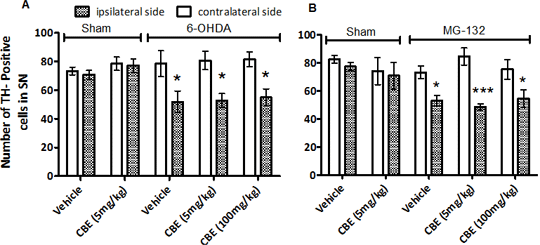
Mutations in the gene for glucocerebrosidase (GBA) not only results in Gaucher’s disease but also increases the risk for Parkinson’s disease (PD) (1). However, GBA mutations do not directly lead to PD, but rather increase the susceptibility for the disease. In rat primary ventral mesencephalic cultures, GBA inhibition by conduritol-β-epoxide (CBE) potentiated neurotoxin-induced dopamine (DA) cell death (2) suggesting an interaction with the processes of cell death in vitro. This study investigated whether GBA inhibition enhances toxin-induced death of DA neurons in the substantia nigra of mice in vivo. C57BL/6 mice were treated daily with CBE (5 or 100 mg/kg, i.p.) or vehicle (0.9% saline) for 10 days. On day 3, 6-hydroxydopamine (6-OHDA; 7.5 µg/4 µl, n=6), MG-132 (0.4 µg/1.5 µl, n=6) or vehicle was injected into the striatum using standard stereotaxic techniques under isoflurane anaesthesia. Mice were culled 14 days after lesioning and the number of tyrosine hydroxylase (TH)-containing cells was determined by immunohistochemistry. A separate group of mice were treated with CBE (5 or 100 mg/kg i.p.) or vehicle for 10 days. GBA activity was determined in brain stem as previously described (3). Data are presented as mean±SEM. Data were analysed by one or two-way ANOVA followed by Newman Keuls or Dunnett’s post-hoc test. CBE significantly inhibited GBA activity in mouse brainstem compared to vehicle-treated controls (vehicle: 54.3±8.6, CBE (5 mg/kg): 15.8±5.8, CBE (100 mg/kg): 13.5±3.8 nmoles/h/mg protein, n=4). Both 6-OHDA and MG-132 reduced the number of TH+ve cells in the SN. CBE (5 mg/kg) did not alter the number of nigral TH+ve cells when given alone. Similarly, CBE (5 or 100 mg/kg) did not alter the loss of dopaminergic neuronal cells following toxin administration (Figure1A and B). Chronic GBA inhibition in mice does not enhance the susceptibility of dopaminergic neurons to oxidative stress or proteasomal inhibition in vivo as previously suggested in in vitro studies (2). The reason for this is unclear as GBA was inhibited by over 70% following 10 days CBE treatment. Further work is required to determine the role of GBA in the increased susceptibility for PD.
Figure 1: Effect of CBE on dopaminergic cell death in (A) 6-OHDA- and (B) MG-132-treated mice. *P<0.05; *** P<0.001 vs. contralateral SN (Newman Keuls) n=6.
(1) Rosenbloom B et al (2011). Blood Cells Mol Dis 46: 95-102. (2) Proceedings of BPS, http://www.pa2online.org/abstracts/Vol3Issue2abst001P.pdf (3) Lee et al (2005). Biochem Biophys Res Commun 337: 701-707.
|


