

| 136P London, UK Pharmacology 2016 |
Cardioprotective effect of neoflavonoid latifolin isolated from dalbergia odorifera on isoproterenol-induced myocardial ischemia in rats
Introduction: The imbalance between coronary oxygen supply and myocardial demand promotes massive generation of reactive oxygen species (ROS)1, which induces to ischemic heart disease (IHD). The present study was designed to evaluate the anti-oxidative stress activity of latifolin in isoproterenol (ISO)-induced rat myocardial ischemia model, and further investigate its underlying mechanism.
Method: Forty male Sprague-Dawley rats were divided randomly into five groups: control group, ISO group, latifolin (100, 50, 25mg/kg/d) treated groups. Rats were treated for five days and the myocardial ischemia model was established by subcutaneous injection (s.c.) of 30 mg/kg of ISO in normal saline daily for last 2 consecutive days2. Lead II electrocardiograph was recorded continually after last administration, and the levels of cardiac troponin I (cTnI), aspartate aminotransferase (AST) in serum and the levels of AST, lactate dehydrogenase (LDH), superoxide dismutase (SOD) and malondialdehyde (MDA) in myocardial tissue were measured using commercial kits. Paraffin sections from heart tissue were stained with hematoxylin-eosin (H&E). Frozen sections from heart tissue were used for the estimation of cardiac reactive oxygen species (ROS) using DCFH-DA probe. The protein levels of nuclear factor erythroid 2-related factor 2(Nrf2), heme oxygenase-1 (HO-1) and NAD(P)H: quinine oxidoreductase (NQO1) in myocardial tissue were measured by Western Blot.
Results: Pretreatment with latifolin inhibited the elevation of ST-segment induced by ISO (Fig. 1). The levels of serum markers (cTnI and AST) were decreased and the levels of myocardium markers (AST and LDH) were increased after latifolin pretreatment (Table 1, 2). And due to the intervention of latifolin, the activity of SOD increased and the level of MDA decreased in myocardial tissue (Table 2). Latifolin also protects myocardium by decreasing the infiltration of inflammatory cells, reducing the degree of interstitial edema and necrosis, which may be due to reducing the generation of ROS. In addition, pretreatment with latifolin promotes the expression of nuclear Nrf2, cytoplasmic Nrf2, HO-1 and NQO1 in myocardial tissue compared to ISO group (Fig.2).
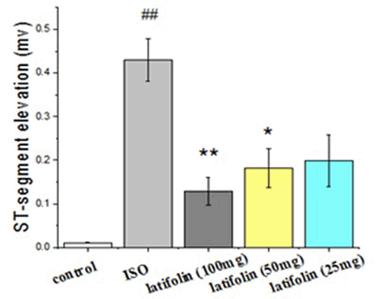
Fig.1. Effect of latifolin on ST-segment changes in ISO-induced myocardial ischemia in rats. Values are mean ±SEM; n=8 in each group. ##p<0.01 vs control group, *p<0.05 vs ISO group, **p<0.01 vs ISO group.
Table 1. Effect of latifolin on cTnI and AST in serum
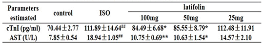
Table 2. Effect of latifolin on AST, LDH, SOD and MDA in myocardial tissue
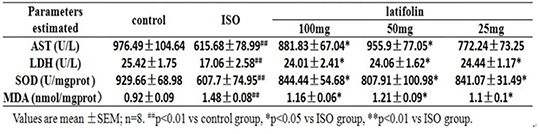
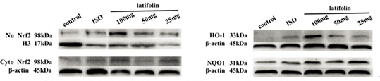
Fig.2 Effect of latifolin on protein expression of Nrf2, HO-1 and NQO1
Conclusion: Pretreatment with latifolin protects myocardium away from ischemic injury. This effect might be relate to the activation of Nrf2 signal pathway which serves as the main molecule mechanism of anti-oxidative stress.
References:
1.Hua Li et al. (2016). Sci Rep 6:23693.
2.Shou-bao Wang, et al. (2009). European Journal of Pharmacology 615:125-132.