

| 160P London, UK Pharmacology 2016 |
Tiron offers protection against H2O2-induced mitochondrial DNA damage
Introduction: Reactive oxygen species (ROS) are metabolically produced free radicals maintained by cellular antioxidants under normal physiological conditions (1). Oxidative stress can occur when ROS production is augmented beyond manageable thresholds. ROS have been implicated in the development of diseases such as Alzheimer’s and chronic kidney disease (1). Studies have been successfully undertaken to investigate the use of antioxidant supplementation to inhibit the effects of ROS in cellular and disease models (2). Tiron, a superoxide scavenger, has been shown to be 100% protective against UV-induced ROS damage in human dermal fibroblasts (3). The aim of this study was to investigate the use of in vitro Tiron supplementation to protect against ROS-induced mitochondrial DNA (mtDNA) strand breaks in human renal proximal tubular (HKC-8) and foetal astrocyte (SVGp12) cell lines.
Method: Cells were pre-treated with Tiron (3mM) for 24 hours prior to induction of ROS damaging hydrogen peroxide (H2O2) at 5mM in HKC-8 and 0.5mM in SVGp12 cells (3). The concentrations of H2O2 used were determined by titration in each cell line to ensure maximal damage was induced. MtDNA strand breaks were assessed by the efficiency of qPCR amplification in a 1kb amplicon following a shorter amplicon (83bp) normalisation (3,4). Data is presented normalised to the cell control and as mean +/- standard deviation (SD), analysed by one-way ANOVA and Dunnett’s posthoc test (p≤0.05).
Results: Pre-treatment with Tiron was found to be 100% protective in preventing mtDNA strand breaks in both HKC-8 (±4%, n=6) (figure 1) and SVGp12 (±2%, n=5) (figure 2) cell lines with induced damage of 9% (±4%) and 10% (±4%) respectively. Additionally, Tiron demonstrated no significant reduction in viability across all cell lines using the TOX-8 assay (5).
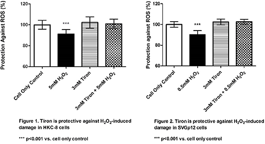
Conclusion: This study indicates the potential use of Tiron to protect against H2O2-induced damage across different clinically relevant cell types. Further investigation is required to assess the protective capabilities of Tiron against natural cellular ROS production, thus establishing a more clinical model. However, the mechanism by which Tiron offers protection requires further elucidation.
References
1. Halliwell B and Gutteridge JMC (2015) Free Radicals in Biology & Medicine. Oxford University Press: Oxford, pp 511-638.
2. Gallego-Villar L et al (2014) Biochemical and Biophysical Research Communications 452: 457-461
3. Oyewole A et al (2013) FASEB Journal 28: 485-494
4. Birch-Machin MA et al (2014) unpublished methodology through personal communication: Newcastle
5. Anoopkumar-Dukie S et al (2005) The British Journal of Radiology 78: 945-947