Print version
Search Pub Med
| 174P London, UK Pharmacology 2016 |
The role of extracellular vesicles as markers and mediators of ANCA vasculitis
Introduction: Systemic vasculitis associated with autoantibodies to neutrophil cytoplasmic antigens (ANCA) is an autoimmune disease which affects blood vessels of all sizes throughout the body1. It can result in life threatening multisystem failure. Currently, vasculitis is underdiagnosed and has a high relapse rate2. This study hypothesised that extracellular vesicles (ECVs) isolated from patients with vasculitis, before and after treatment, would play differing functional roles when applied to murine bone marrow derived macrophages by activating a defined inflammatory response in vitro and so represent a potential new therapeutic option.
Method: Samples were analysed from healthy volunteers and patients with vasculitis at first presentation to the treating clinician (pre-treatment) and in treatment-induced remission (post-treatment). ECVs were isolated from plasma samples and quantified to establish the size of particles present in the samples. The vesicles were then added to mouse bone marrow derived macrophages (BMDMΦ). Uptake was assed using flow cytometry and mRNA expression was measured by qPCR. Data are presented as mean±SD. Statistical significance was calculated using a one way ANOVA with Bonferroni’s multiple comparison post-test for comparison to healthy controls and for all FLOW analysis. For pre and post treatment, a paired T test was used.
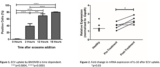
Results: To ascertain the period of time required for uptake, fluorescently labelled vesicles were incubated with BMDMΦ. Analysis by flow cytometry (Figure 1) confirmed the greatest uptake by cells was 18 hours after ECV addition (80.3% ± 1.0% percentage positive cells). ECV uptake was confirmed by immunocytochemistry which indicated that the labelled ECVs were present intracellularly. After uptake was established, ECVs from vasculitis patients were added to BMDMΦ to examine the differences in inflammatory gene expression. Murine macrophages with ECVs from patients post-treatment produced a statistically significant increase in the mean expression of Il10 (Figure 2), encoding IL-10 (an anti-inflammatory cytokine) by 0.645 fold (n=5, p=0.03).

Conclusion: ECVs isolated from plasma samples from patients with vasculitis can be taken up my murine macrophages and induce an anti-inflammatory response in vitro. Further assessment should determine the vesicle components that elucidate these phenotypic changes.
References:
1.Janette JC et al. (2012). Clinical and Experimental Nephrology 17: 603-606
2. Flossmann, O et al. (2013). Annals of the Rheumatic Diseases 70: 488-494

