
Print version
Search Pub Med

| 201P London, UK Pharmacology 2016 |
Relative role of COX-1 and COX-2 in renal versus systemic vascular responses
Introduction: Non-steroidal anti-inflammatory drugs (e.g. ibuprofen) are amongst the most widely used drugs worldwide and act by inhibition of cyclo-oxygenase (COX)-2. Their use is associated with increased risk of heart attacks and strokes. The mechanisms for this cardiovascular toxicity remain uncertain with two major hypotheses proposed: (1) that COX-2 is expressed throughout the systemic vasculature where it mediates production of cardioprotective prostacyclin or (2) that COX-2 is localised to the kidney where it exerts powerful indirect regulatory effects on the cardiovascular system. Here we have compared the contribution of COX isoforms to vascular responses in large systemic arteries versus renal arterioles to better understand how COX-2 inhibition produces cardiovascular dysfunction.
Methods: Aorta and kidney were isolated from wild-type C57Bl/6 mice (8-10wks, n=4-5 animals for all experiments). Vascular responses in Kreb’s buffer were measured in aorta using wire myography and in renal arterioles by imaging of 150μm live tissue slices. Vessels were pre-treated with diclofenac (3μM), valdecoxib (3μM) or SC-560 (100nM). After 10 mins, concentration response curves to phenylephrine were recorded. Other vessels/slices were pre-treated with diclofenac, pre-contracted phenylephrine (EC80 concentration) then stimulated with treprostinil (0.1-10μM). Prostacyclin (measured as 6-keto-PGF1α) release from A23187 (50μM)-stimulated aorta/kidney was measured by immunoassay. Data are given as mean±SEM for n independent experiments and considered statistically (*) where p<0.05 by two-way ANOVA.
Results: In the aorta, combined COX-1+COX-2 inhibition with diclofenac had no effect on contractile responses to phenylephrine (Figure 1a), however, in renal arterioles, diclofenac increased the sensitivity of vessels to phenylephrine-induced contraction (Figure 1b). This effect was reproduced with a specific COX-2 inhibitor (-logIC50 vehicle: 5.6±0.2; valdecoxib: 6.2±0.1; p=0.02) whereas a specific COX-1 inhibitor had no effect (-logIC50 vehicle: 5.7±0.4; SC-560: 5.7±0.4; p=0.85). Aorta and kidney had a similar capacity to generate prostacyclin (Figure 1c), however, when an exogenous prostacyclin analogue (treprositinil) was added to vessels it produced relaxation only of renal arterioles (Figure 1d).
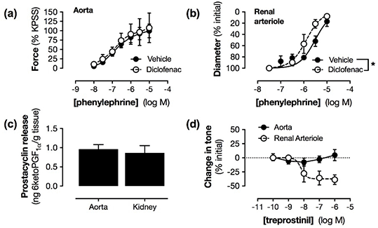
Figure 1: Contribution of COX and prostacyclin to vascular function in aorta versus renal arterioles.
Conclusions: These data demonstrate the existence of a COX-2-prostacyclin-vasodilation axis in the kidney which is not present in large systemic vessels. This may reflect relative differences in COX expression patterns and sensitivity of different vascular beds to prostacyclin. These data add to support to the idea that cardiovascular side effects of COX-2 inhibitors arise in the kidney.