Print version
Search Pub Med
| 008P London, UK 8th European Workshop on Cannabinoid Research |
2-arachidonoylglycerol favors myelin debris phagocytosis by microglia supporting remyelination
Introduction: Remyelination is a regenerative mechanism following demyelination that involves the mobilization and differentiation of oligodendrocyte progenitor cells (OPCs); nevertheless, this process is incomplete or fails in multiple sclerosis (MS)1. Microglia exert a phenotype associated with myelin debris phagocytosis and OPC recruitment during demyelination2,3. In a progressive model of MS, the Theiler's murine encephalomyelitis virus-induced demyelinating disease (TMEV-IDD), we have previously shown that there is a prominent demyelination in the corpus callosum (CC) at 35 days post-infection (dpi), followed by incomplete remyelination at 60 dpi that is abolished as the disease progresses4. The aim of this study was to investigate whether the endocannabinoid 2-arachidonoylglycerol (2-AG) could target microglia to favor myelin debris phagocytosis promoting the endogenous reparative process of remyelination.
Methods: Adult female SJL/J mice were infected with TMEV virus (DA strain). At 28 dpi, 2-AG or vehicle were administered for 7 or 14 days with subcutaneous miniosmotic pumps (Alzet, 3,5 mg/kg) to study microglia dynamics in the CC by Iba1 immunohistochemistry at 35, 42 and 60 dpi. In a subset of in vitro experiments using microglia cultures from postnatal P0-P2 rat brains, we studied markers of phagocytosis by RT-PCR and performed a functional assay with Cy3-stained myelin (2 μg/ml). Data are given as mean ± SEM (n animals) and analysis was performed using one-way ANOVA followed by non-parametric tests (U-Mann Whitney and Kruskall Wallis), as applicable.
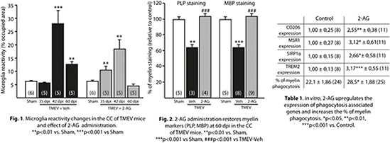
Results: 2-AG-treated mice display anticipated microglial activation (% of Iba1 occupied area) associated with myelin phagocytosis in the CC at 35 dpi and accelerated resolution of inflammation at 60 dpi compared to vehicle (Figure 1), an effect that is accompanied by the restoration of myelin markers (PLP, MBP) to control levels (Figure 2). In vitro experiments show that 2-AG (1 nM) treated microglia increases the expression of phagocytosis associated genes like CD206, MSR1, SIRP1α and TREM-2, and the % of myelin engulfment (Table1).
Conclusions: 2-AG induces a phagocytic activation state in microglia improving myelin debris clearance and contributing to a pro-regenerative milieu for OPC migration and differentiation in the damaged CC.
References:
1. Goldschmidt T et al. (2009). Neurology 72:1914-1921.
2. Jurevics H et al. (2002). J Neurochem 82:126-136.
3. Olah M et al. (2012). Glia 60:306-321.
4. Mecha M et al. (2013). Exp Neurol 250:348-52.


