
Print version
Search Pub Med

| 031P London, UK Pharmacology 2017 |
The effect of a phosphoinositide 3-kinase-α inhibitor in vascular inflammation.
Introduction: Cardiovascular inflammation is associated with endothelial cell (EC) damage, resulting in leukocyte trafficking and oedema formation (1). Inflammation disrupts EC junctions, increasing microvascular permeability, and resulting in a positive-feedback loop of inflammatory events. The phosphoinositide 3-kinase, PI3Kα, stimulates this endothelial response (2) and this study examined the actions of BYL-719, a PI3Kα selective inhibitor, on inflammatory responses.
Method: Immunofluorescence and confocal imaging were used to investigate actions of TNF-α in the presence and absence of BYL-719 on human vein umbilical endothelial cells (HUVEC). In vivo analysis was carried out per the UK Home Office Animals (Scientific Procedures) Act 1986. CD1 mice were anaesthetised using isoflurane, to test BYL-719 in a zymosan-induced model of dorsal skin inflammation on neutrophil accumulation (measured by myeloperoxidase) and oedema formation (measured by Evans Blue accumulation) (3). Data were analysed using 1-way or 2-way ANOVA with post-hoc Bonferonni\\\\\\'s test.
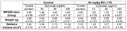
Results: BYL-719 significantly reduced TNF-α-induced EC shape changes, including cell area and elongation (Table 1), impeding in vitro cytokine-induced inflammation. Whilst there was no effect on neutrophil accumulation in vivo, there was significant reduction in weight of treated areas and oedema formation was inhibited by BYL-719 (Table 2).
Table 1: Effect of BYL-719 in vitro on cell morphology. HUVECs treated with TNF-α for 16-18 hr, followed by 1 hr inhibitor treatment at 1, 3 and 10 μM and examined via immunofluorescence. N=3 independent experiments in duplicates, 3 images/condition and 10 cells/image; p* < 0.05 between control and TNF-α groups.
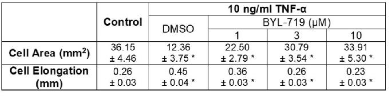
Table 2: Effect of BYL-719 in vivo on MPO/Protein levels, tissue weight and oedema formation. Mice were pre-treated with BYL-719 intraperitoneally for 30 min, intradermally injected with zymosan (50 μl/site) for 4 hr, followed by ex vivo MPO assay, weight and oedema volume measurements. Oedema was quantified by measuring three planes of view: width(x), height(y) and depth(z) from each of the intradermal sites; volume of oedema = ((π/6)xyz). N=3 independent experiments in duplicates; p* < 0.05 between Tyrode solution vs zymosan groups.
Conclusions: Our findings show that the PI3Kα inhibitor, BYL-719, reduces endothelial activation and inhibits inflammatory oedema formation. We conclude that there is a potential for PI3K inhibitors to act as anti-oedema agents in cardiovascular-related inflammatory conditions. We thank the British Heart Foundation (BHF) for funding this work.
References:
1) Pearson et al., 2003. Circulation. 107:499-511.
2) Cain et al., 2010. J Cell Biol. 188:863-876.
3) Sawyer et al., 2011. PLoS One. 6:e14671.