| 156P London, UK Pharmacology 2017 |
Opioids disinhibit midbrain periaqueductal grey neurons projecting to the rostral ventral medulla. Direct validation of the disinhibition hypothesis.
Introduction: Projections from the midbrain periaqueductal grey (PAG) to the rostral ventral medulla (RVM) form an important component of the descending analgesic pathway, which when activated can inhibit ascending pain signals1. Opioids are potent analgesics thought to activate this descending pathway by inhibiting GABAergic interneurons that tonically inhibit PAG projection neurons2. Whilst widely accepted, evidence supporting this disinhibition hypothesis is largely indirect. Most previous reports have been conducted on unidentified PAG neurons and the origin of the GABAergic tone is unknown. The aim of this study was to specifically investigate the effect of opioids on the input and activity of PAG projection neurons.
Methods: Rhodamine-conjugated microspheres were stereotaxically injected into the RVM of anesthetised Sprague Dawley rats (3-4 weeks old) to retrogradely label PAG projection cells. In addition, some animals also received the channelrhodopsin construct, AAV5-hSyn-ChR2(H134A)-eYFP which was injected into the PAG. Brain slices were prepared 5 (retrograde only) or 35 (retrograde + ChR2) days post-surgery and in vitro whole cell voltage clamp recordings of postsynaptic currents were conducted from rhodamine labelled PAG neurons. The effects of mu- (DAMGO, 3μM), delta- (deltorphin-II, 300nM) and kappa- (U69593, 300nM) opioid receptor agonists were studied on optically (475 nm) or electrically evoked responses. Data are normalised to baseline values and given as mean ± S.E.M. Analysis was performed using Student’s paired or unpaired t-tests as appropriate.
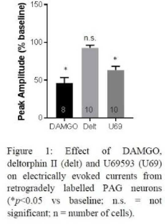
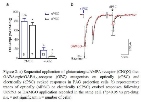
Results: Both DAMGO and U69593 reduced electrically evoked inhibitory currents in PAG projection cells (Figure 1) and increased the paired pulse ratio (DAMGO: 117.0 ± 5.4%, n=8; U69593: 112.6 ± 5.1%, n=10; both p<0.05), indicating a presynaptic mechanism. In contrast, deltophin-II had no effect. When local inputs were evoked optically, most responses were inhibited by the GABAA receptor antagonist gabazine, whilst AMPA receptor antagonists had no significant effect (Figure 2a). In addition, preliminary findings (n=3) suggest these optically evoked currents are inhibited predominantly by DAMGO whilst U69593 had little effect (Figure 2b).
Conclusion: Our findings offer direct support for the disinhibition hypothesis. Optogenetic control of local inputs reveal these to be predominantly GABAergic indicating local interneurons provide significant inhibitory input to PAG projection neurons. Our data suggest that whilst overall inhibitory input to PAG projection cells is sensitive to both mu- and kappa- agonists, local interneurons are particularly susceptible to mu-opioid receptor control.


References:
1 Ossipov, M. H. et al.(2010) J Clin Invest 120, 3779-3787.
2 Lau, B. K. et al.(2014) Curr Opin Neurobiol 29, 159-164.

