Print version
Search Pub Med
| 174P London, UK Pharmacology 2017 |
Molecular imaging of I2-imidazoline receptors (I2R): in vitro binding of [3H]BU99008 across species, animal models and disease
Introduction: The function of the I2-Imidazoline receptors (I2R) is largely unknown, but evidence shows involvement in brain disorders, including depression and Alzheimer’s disease [1]. I2R were found to be expressed in astrocytes and upregulated in human glioma and rodent gliosis. Radiolabelled I2R ligands, such as ([3H/11C]BU99008, are therefore potential astrocytic biomarkers. To investigate the suitability of BU99008 as astrocyte marker, we employed [3H]BU99008 in preliminary studies across species, different animal models that include astrocyte activation and human post-mortem tissue from patients with Alzheimer’s disease (AD) and healthy controls.
Methods: In vitro autoradiography was performed to investigate and quantify binding pattern in brain tissue from the following animals and models: 1) mouse model of AD [5xFAD and respective wild-types (WT)]; 2) mouse model of glioblastoma; 3) rat model of Traumatic Brain Injury (TBI) and 4) post-mortem tissue from patients with dementia. Fresh-frozen sections were incubated with 2 nMol/L [3H]BU99008. Binding pattern was assessed and quantified in selected brain regions. Staining for Amyloid β and GFAP was performed in selected sections for comparison.
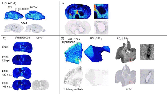
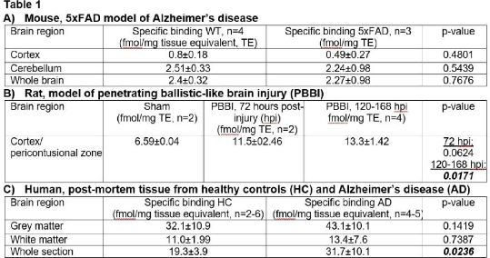
Results: Specific binding in the mouse whole brain, cortex and cerebellum was found to be very low for both WT and 5xFAD mice, without significant differences (Table 1A/Figure 1A). In a mouse model of glioblastoma, binding was remarkably absent in the centre of the tumour but accumulation was observed in the surrounding tissue, coinciding with GFAP staining (Table 1B/Figure 1B). Binding in rats was slightly higher as compared to the mouse brain (Table 1C/Figure 1C). Using a penetrating injury, binding in the pericontusional zone was non-significantly increased at 72 and significantly increased at 120-168 hours post injury as compared to sham-operated animals. In human post-mortem tissue (Table 1D/Figure 1D), binding was much more pronounced, almost exclusively attributed to grey matter. In the whole section, a significant 64% increased binding was found for AD cases, likely attributed to grey matter (+34%). Binding seemed to co-localize with staining for amyloid β and GFAP.
Conclusion: [3H]BU99008 binding in the rodent brain was found to be low and not very indicative of astrocyte activation, when compared to immunohistochemical staining. In contrast, human post-mortem brain sections showed a much higher general binding which co-localized well with GFAP staining for astrocytes. Further studies are needed to dissect the changes of I2R in relation to astrocyte activation.
References:
1. Garcia-Sevilla JA et al. (1999). Ann N Y Acad Sci 881: 392-409.


